Figures & data
Figure 1 Combined treatment with GSK126 and Gefitinib exhibited synergic effect on survival fraction in different types of cancer cells.
Notes: The cells were incubated in various concentrations of GSK126 (0.39–12.50 µM) and Gefitinib (0.26–8.30 µM) or combinations of both (1:1) for 48 hours. Dose– response curves of human cancer cell lines treated with Gefitinib or GSK126 alone or in combination: (A) MGC803, (B) A549, (C) PC-3, (D) MDB-231.
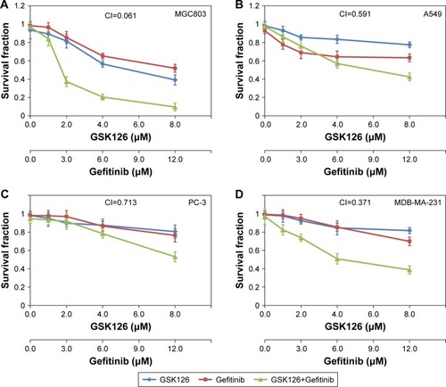
Figure 2 Combined treatment with GSK126 and Gefitinib caused enhanced apoptosis.
Notes: (A) Analysis of apoptosis by Annexin V-FITC/propidium iodide (PI) double staining of MGC 803 cells after 48 hours of treatment with 8.30 µM Gefitinib and/or 12.50 µM GSK126. (B) Quantification of the number of apoptotic cells in (A). *P<0.05, n=3.
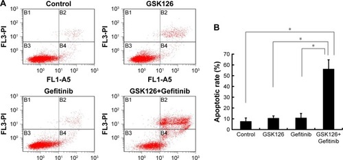
Figure 3 Combined treatment with GSK126 and Gefitinib had synergic effect on autophagy induction.
Notes: (A) Representative blots of ULK and LC3 in MGC803 cell treated with 8.30 µM Gefitinib and/or 12.50 µM GSK126 for 48 hours. (B) Quantitative analysis of p-ULK/ULK and LC3-II/LC3-I. (C) MGC 803 cells were pre-treated with 1 mM 3-MA for 1 hour and incubated with Gefitinib and/or GSK126 at the indicated concentration for 48 hours. Western blotting analysis was conducted for p-ULK/t-ULK and LC3-II/LC3-I. (D) Propidium iodide analysis was conducted for apoptotic cells after 3-MA pretreatment. *P<0.05, **P<0.01.
Abbreviations: 3-MA, 3-methyladenine; p-ULK, phosphorylated ULK; ULK, Unc-51-like autophagy activating kinase; t-ULK, total ULK.
Abbreviations: 3-MA, 3-methyladenine; p-ULK, phosphorylated ULK; ULK, Unc-51-like autophagy activating kinase; t-ULK, total ULK.
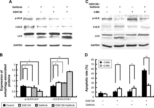
Figure 4 Combined treatment with GSK126 and Gefitinib resulted in greater inhibition of mTOR pathway.
Notes: MGC803 cells treated with 8.30 µM Gefitinib and/or 12.50 µM GSK126 for 48 hours. (A) Representative blots of phosphorylation of Akt, mTOR, and S6 by Western blotting analysis. Quantitative analysis of p-Akt/t-Akt (B), p-mTOR/t-mTOR (C), and p-S6/t-S6 (D) was demonstrated. Combination treatment with GSK126 and Gefitinib caused a greater decrease in p-Akt/t-Akt, p-mTOR/t-mTOR, and p-S6/t-S6. *P<0.05, **P<0.01.
Abbreviations: mTOR, mammalian target of rapamycin; p-mTOR, phosphorylated mTOR.
Abbreviations: mTOR, mammalian target of rapamycin; p-mTOR, phosphorylated mTOR.
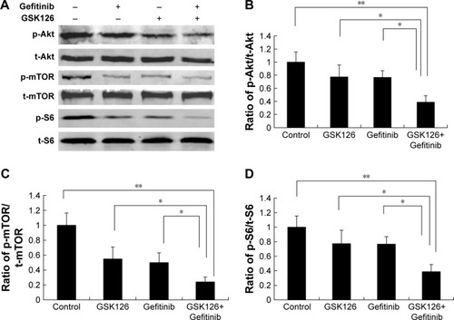
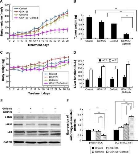
Figure 5 Combined treatment with GSK126 and Gefitinib significantly inhibited tumor xenograft in nude mice but did not affect body weight and liver function.
Notes: The nude mice transplanted with MGC803 cells tumor xenografts were randomly divided into four groups that received i.p. injection of GSK126 (200 mg/kg) and/or Gefitinib (30 mg/kg) for 4 weeks. (A) Tumor volume was recorded every other day. (B) Mice were sacrificed on day 28 and tumor wet weight was measured. (C) The body weight of nude mice was recorded every other day. (D) The content of AST and ALT was determined by ELISA using commercial kits. (E) Representative blots of ULK and LC3 proteins in tumor tissues. (F) Quantitative analysis of p-ULK/t-ULK and LC3-II/LC3-I. *P<0.05, **P<0.01, n=8–10 mice/group.
Abbreviations: i.p., intraperitoneal; p-ULK, phosphorylated ULK; ULK, Unc-51-like autophagy activating kinase; ALT, alanine transaminase; AST, aspartate transaminase; t-ULK, total ULK.
Abbreviations: i.p., intraperitoneal; p-ULK, phosphorylated ULK; ULK, Unc-51-like autophagy activating kinase; ALT, alanine transaminase; AST, aspartate transaminase; t-ULK, total ULK.