
Figures & data
Table 1 The sequences of primers used in real-time PCR
Figure 1 Cytotoxicity of fenofibrate, paclitaxel, TRAIL, ABT-737, and doxorubicin in human breast cancer cells.
Notes: (A) Fenofibrate inhibited human breast cancer cell growth in vitro. The cancer cells were incubated in the presence of various concentrations of fenofibrate for 24 and 48 hours. Cell viability was determined by the MTT assay. Each point represents the mean of the data of three independent experiments; bars represent SD; *P<0.05 vs control; **P<0.01 vs control. (B–E) Paclitaxel, TRAIL, ABT-737, and doxorubicin suppressed human breast cancer cell growth in vitro. The cancer cells were incubated in the presence of various concentrations of fenofibrate for 24 hours. Cell viability was determined by the MTT assay. Each point represents the mean of the data of three independent experiments; bars represent SD; *P<0.05 vs control; **P<0.01 vs control.
Abbreviations: DOX, doxorubicin; TRAIL, tumor necrosis factor-related apoptosis-inducing ligand.
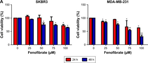
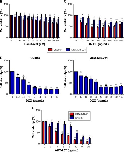
Figure 2 Fenofibrate potentiates chemosensitivity to human breast cancer by modulating apoptosis.
Notes: (A) SKBR3 cells treated with indicated concentrations of fenofibrate alone and/or combined with indicated concentrations of paclitaxel, TRAIL, ABT-737, and doxorubicin for 24 hours. Cell viability was determined by the MTT assay. Each point represents the mean of the data of three independent experiments; bars represent SD; *P<0.05 vs control; **P<0.01 vs control. (B, C) SKBR3 cells treated as A without doxorubicin treatment. After that, cells were subjected to Annexin-V-FITC and PI staining. Flow cytometry assay was performed to detect the percentage of apoptotic cells. Data are presented as mean ± SD, n=3. **P<0.01 vs control. (D) MDA-MB-231 cells treated with indicated concentrations of fenofibrate alone and/or combined with indicated concentrations of paclitaxel, TRAIL, ABT-737, and doxorubicin for 24 hours. Cell viability was determined by the MTT assay. Each point represents the mean of the data of three independent experiments; bars represent SD; *P<0.05 vs control; **P<0.01 vs control. (E, F) MDA-MB-231 cells treated as D without doxorubicin treatment. **P<0.01 vs control. SKBR3 cells (G) were treated with indicated concentrations of fenofibrate and TRAIL, and MDA-MB-231 cells (H) were treated with indicated concentrations of fenofibrate and ABT-737. MTT assay was performed and the data were analyzed by Chou-Talalay method (combination index >1 indicates antagonism, =1 indicates additivity, and <1 indicates synergy).
Abbreviations: DOX, doxorubicin; TRAIL, tumor necrosis factor-related apoptosis-inducing ligand; PI, propidium iodide; FENO, fenofibrate; PAC, paclitaxel; FITC, fluorescein isothiocyanate.
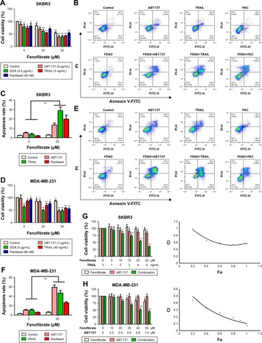
Figure 3 Treatment with fenofibrate resulted in downregulation of Mcl-1 and Bcl-xl and upregulation of Bok and Bax.
Notes: (A) SKBR3 and MDA-MB-231 cells treated with indicated concentrations of fenofibrate and combined with indicated concentrations of paclitaxel, TRAIL, and ABT-737 for 24 hours. After that, PARP and caspases proteins were assessed by Western blotting assay. (B) SKBR3 and MDA-MB-231 cells treated with indicated concentrations of fenofibrate for 24 hours. After that, Bcl-2 family proteins were assessed by Western blotting assay. (C) SKBR3 and MDA-MB-231 cells treated with indicated concentrations of fenofibrate for 24 hours. After that, qRT-PCR analyzed expression level of Bcl-2 family proteins. Data were presented as mean ± SD for three independent experiments. *P<0.05 vs control; **P<0.01 vs control.
Abbreviations: TRAIL, tumor necrosis factor-related apoptosis-inducing ligand; PARP, poly (ADP-ribose) polymerase 1; caspase, the cysteine-aspartic acid protease.
Figure 4 The intrinsic apoptotic pathway is initiated in the sensitization effect of fenofibrate on ABT-737, TRAIL, and paclitaxel in human breast cancer cells.
Notes: SKBR3 (A) and MDA-MB-231 (B) cells were treated as mentioned in . After treatment, the cells were stained with JC-1 and subjected to flow cytometry assay to detect the mitochondrial outer membrane permeabilization. Data are presented as mean ± SD, n=3. **P<0.01 vs control.
Abbreviations: TRAIL, tumor necrosis factor-related apoptosis-inducing ligand; JC-1, 5,5′,6,6′-tetrachloro-1,1′,3,3′-tetraethylbenzimidazolylcarbocyanine iodide; FENO, fenofibrate; PAC, paclitaxel.
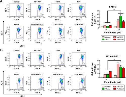
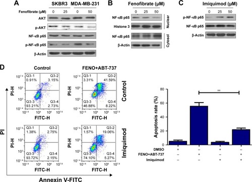
Figure 5 Fenofibrate exerted its chemo-sensitization effects in human breast cancer cells apoptosis via activation of AKT/NF-κB pathway.
Notes: (A) SKBR3 and MDA-MB-231 cells treated with indicated concentrations of fenofibrate for 24 hours. After that, AKT and NF-κB proteins were assessed by Western blotting assay. (B) Nuclear translocation of NF-κB in MDA-MB-231 cells was determined by Western blotting after treatment with indicated concentrations of fenofibrate for 24 hours. (C) Activation of NF-κB in MDA-MB-231 cells was determined by Western blotting after treatment with indicated concentrations of imiquimod. (D) MDA-MB-231 cells were pretreated with 50 µM imiquimod for 24 hours followed by co-treatment with 25 µM fenofibrate and 1 µg/mL ABT-737 for another 24 hours. Flow cytometry assay was used to detect the cell apoptosis. Data were presented by mean ± SD for three independent experiments. **P<0.01 vs control.
Data availability
All data for this study are presented in this published article.