Figures & data
Figure 1 Effects of VEGF-A and Ang-2 siRNAs in Ishikawa cells.
Notes: (A) Relative VEGF-A mRNA expression was analyzed by real-time PCR. (B) Relative Ang-2 mRNA expression. (C) Relative VEGF-A protein expression. (D) Relative Ang-2 protein expression. (E) Western blotting analysis of protein expression in Ishikawa cells after transfection. **P<0.01.
Abbreviations: GAPDH, glyceraldehyde 3-phosphate dehydrogenase; siRNA, small interfering RNA; VEGF, vascular endothelial growth factor.
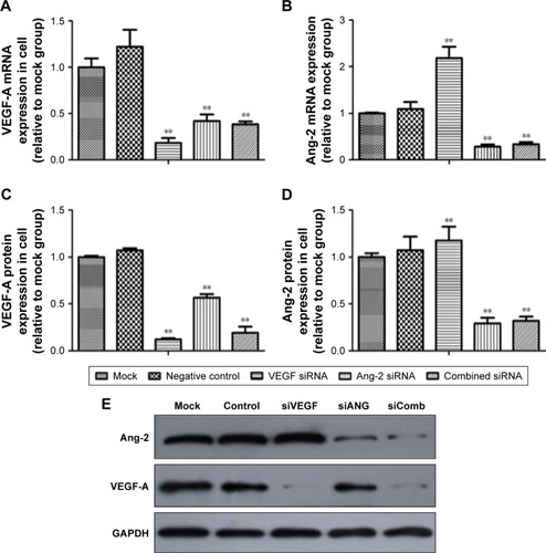
Figure 2 VEGF-A and Ang-2 siRNAs inhibited proliferation and invasion abilities of Ishikawa cells.
Notes: (A) MTT proliferation in Ishikawa cells. (B) Representative images of the wound-healing assay showed the occlusion of the artificial wound performed in the post-transfected 24 hours after wounding. (C and D) Transwell assay demonstrated the suppressed invasive ability of post-transfected cells. The average number of cells was counted from five random microscopic fields (×200). *P<0.05, **P<0.01.
Abbreviations: siRNA, small interfering RNA; VEGF, vascular endothelial growth factor.
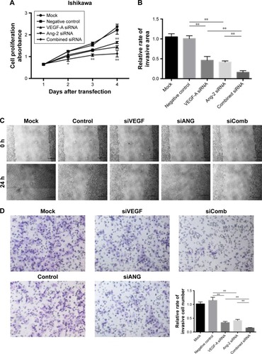
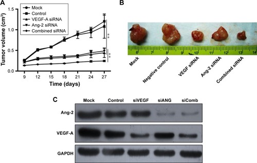
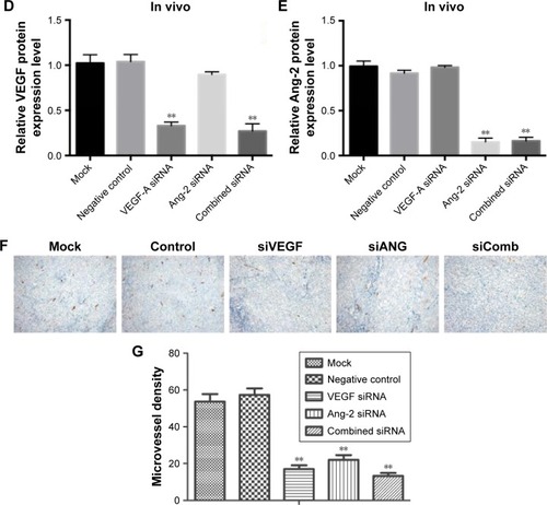
Figure 3 Effects of siRNA therapy on tumor in vivo.
Notes: (A) Tumor growth curve. (B) Tumor volume. (C) Western blotting of VEGF-A and Ang-2 protein expression after siRNA therapy. (D) Analysis of VEGF-A expression in tumor tissues. (E) Analysis of Ang-2 expression in tumor tissues. **P<0.01. (F) Photomicrograph of immunohistochemical staining of vWF (200×). (G) MVD. The arrows are directed to the vessels for MVD. **P<0.01.
Abbreviations: GAPDH, glyceraldehyde 3-phosphate dehydrogenase; MVD, microvessel density; siRNA, small interfering RNA; VEGF, vascular endothelial growth factor; vWF, Von Willebrand factor.