
Figures & data
Figure 1 Brain MRI scans.
Notes: MRI before treatment (A–D): (A) axial T2W shows a hyperintense signal alteration in the right side of the pons; (B) axial T1W post-contrast; (C) sagittal T1W post- contrast; and (B and C) increased enhancement within the tumor. (D) 3D-ASL CBF shows increased CBF value in the right pons. MRI after treatment (E–H): (E) axial T2W shows a decreased hyperintense signal in the right side of the pons compared to pretreatment; (F) axial T1W post-contrast; (G) sagittal T1W post-contrast; and (F and G) disappeared right pontine lesion. (H) 3D-ASL CBF shows decreased CBF value in the right pons compared to pretreatment.
Abbreviations: 3D-ASL, three-dimensional arterial spin labelling; CBF, cerebral blood flow; T1W, T1 weighted; T2W, T2 weighted.
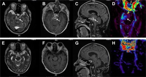
Figure 2 Histopathologic and immunohistochemical examinations of the biopsy tissue.
Notes: (A) H&E stain showed features of anaplastic astrocytoma (WHO grade III) with increased cellularity, nuclear atypia, and mitotic activity. (B) MiB1 (Ki-67) immunohistochemistry showed high proliferative activity with 60% positive cells. (C) GFAP immunohistochemistry. (D) Oligo-2 immunohistochemistry. (E) VEGFR-1 immunohistochemistry. (F) VEGFR-2 immunohistochemistry. (G) CD117 (c-kit). (H) H3K27M immunohistochemistry. Staining was positive for GFAP, oligo-2, VEGFR1, and VEGFR2 and negative for c-kit and H3K27M. Magnification 400×.
Abbreviation: VEGFR, vascular endothelial growth factor receptor.
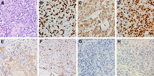
Table 1 Results of molecular tests
Table 2 Survival data of histologically diagnosed brainstem gliomas in adults
Table 3 Case reports of adult BSG treated with antiangiogenic agents