
Figures & data
Figure 1 Schematic representation of the structure of the CRYAB protein (including the N-terminal domain, the flexible C-terminal domain, the WDPF domain, and the α-Crystallin protein domain, and the serine (S) phosphorylation site).
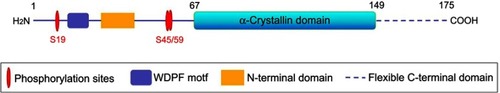
Figure 2 Schematic diagram of CRYAB protein involved in the regulation of apoptosis (inhibition: ┤, activation: →).
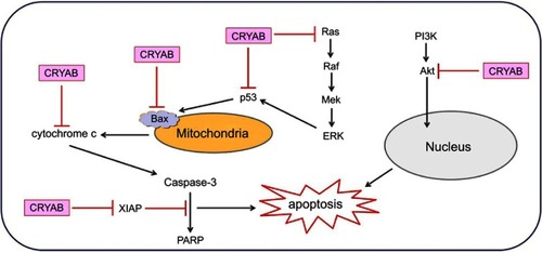
Table 1 Expression and functional characterization of CRYAB in cancer