Figures & data
Figure 1 H&E staining and immunohistochemical staining of specimens in the case. (A) The mass was irregular and infiltrated the adjacent adipose tissue (magnification, x40). (B) the spindle cells were arranged in parallel and partially in a wavy configuration around the mammary duct (magnification, x100). (C) The spindle cells were poorly circumscribed with eosinophilic, pale cytoplasm, sparse nucleus chromatin, and unobvious nucleoli (magnification, x200). (D) Immunohistochemical stain for β-catenin showed nuclear positivity.

Figure 2 Contrast enhanced magnetic resonance imaging of the breast. (A) Before treatment, a well-defined mass (arrow) was observed at the rear of right breast nipple which showed homogeneous strengthening on enhanced magnetic resonance imaging. (B) Five days after the first high intensity focused ultrasound HIFU ablation, a non-contrast enhancement region (arrow) showed up in the previous mass area. (C) Five days after the second treatment, the non-contrast enhancement region was enlarged with irregular boundary and garland-like peripheral enhancement (arrow). (D) Three months after the last treatment, the mass was well ablated and only demonstrated a patchy, heterogeneous architectural distortion region. The nipple retraction was improved.
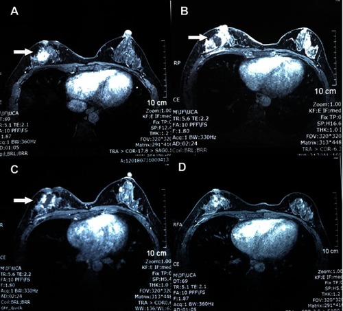
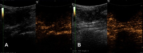
Figure 3 The contrast enhanced ultrasonography images before (A) and 48 minutes after treatment (B). (A) The left image shows a 3.0 cm irregular, hypoechoic mass with obscure boundary at right breast, located beside the nipple. The right image shows the blood flow signal inside the mass 52 seconds after contrast injection. (B) The left image shows the echo of mass enhanced; the right image shows non-contrast enhancement in the mass because of the vessel occlusion caused by high intensity focused ultrasound ablation.