Figures & data
Table 1 Association Between The Expression Of miR-204-3p And Clinicopathologic Feature In Bladder Cancer Patients
Figure 1 miR-204 is downregulated in bladder cancer. (A) The miR-204 expression in bladder cancer tissues compared with that in adjacent non-tumoral tissues was assessed by qRT-PCR. (B) The miR-204 expression in bladder cancer cell lines (HT-1197, HT-1376, J82, RT4, T24, 5637) compared with that in normal human bladder epithelial SV-HUC-1 cells was assessed by qRT-PCR. *P<0.05, **P<0.01.
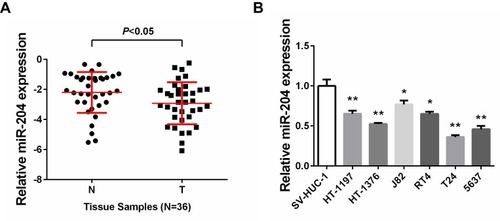
Figure 2 miR-204 inhibits cell growth of bladder cancer cells. (A) 5637 and HT-1376 cells were transfected with miR-204 mimics or inhibitor, respectively. After transfection, qRT-PCR was performed to examine the expression of miR-204. (B) Cell viability was measured by CCK-8 Assay at 0, 24, 48, 72 and 96hrs after miR-204 mimics or inhibitor transfection. (C) Crystal violet staining assay was conducted to evaluate cell proliferation after miR-204 mimics or inhibitor transfection. The quantitative results were shown in right panel. *P<0.05, **P<0.01, ***P<0.001.
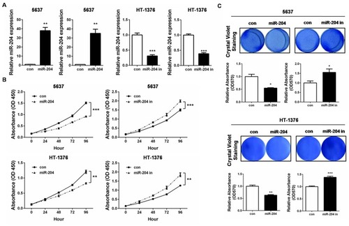
Figure 3 miR-204 inhibits cell migration and invasion of bladder cancer cells. (A) Cell migration ability was investigated with wound healing assay and the images were taken at 0 and 72hrs after miR-204 mimics or inhibitor transfection (40×). (B) Cell invasion ability was investigated with transwell assay and the images were taken at 72hrs after miR-204 mimics or inhibitor transfection (100×). Representative images are shown (left panel). The quantitative results represent the mean±SD in triplicate using the bar graph (right panel). **P<0.01, ***P<0.001.
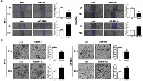
Figure 4 ROBO4 is a direct target of miR-204. (A) Bioinformatics analysis predicts that ROBO4 is a putative target gene of miR-204. The luciferase reporter plasmid containing ROBO4 3ʹ UTR (WT, in green) and ROBO4 3ʹ UTR (MUT, in red) was generated. (B) Luciferase activity assay was performed in 5637 and HT-1376 cells co-transfected with miR-204 mimics and a dual-luciferase reporter containing the ROBO4 3ʹ UTR (WT) and ROBO4 3ʹ UTR (MUT). (C) The expression of ROBO4 in 5637 and HT-1376 cells 48hrs after transfection with miR-204 mimics or controls was measured by qRT-PCR. (D) The expression of ROBO4 in 5637 and HT-1376 cells after 48hrs transfection with miR-204 mimics or controls was measured by Western blot analysis. **P<0.01, ***P<0.001.
Abbreviations: WT, wild-type; MUT, mutant.
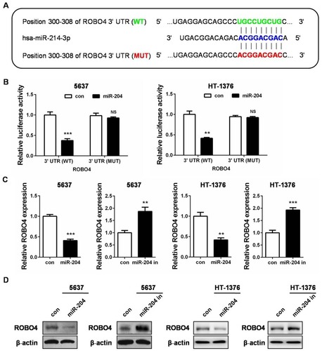
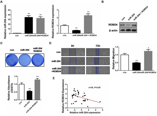
Figure 5 miR-204 mediates a tumor-suppressing role through ROBO4 in bladder cancer. (A) The expressions of miR-204 and ROBO4 in 5637 cells 48hrs after transfection with miR-204 mimics without or with ROBO4 plasmids were measured by qRT-PCR. (B) The expressions of miR-204 and ROBO4 in 5637 cells 48hrs after transfection with miR-204 mimics without or with ROBO4 plasmids were measured by Western blot analysis. (C) Crystal violet staining assay was conducted to evaluate cell proliferation after miR-204 mimics without or with ROBO4 plasmids transfection. The quantitative results were shown in panel below. (D) Cell migration ability was investigated with wound healing assay and the images were taken at 0 and 72hrs after miR-204 mimics without or with ROBO4 plasmids transfection (40×). (E) miR-204 and ROBO4 expression levels exhibited negative correlation. **P<0.01.