Figures & data
Figure 1 Chemical structure of apiosporamide.
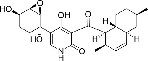
Table 1 The Primers Of Real-Time PCR
Table 2 IC50 Values (Mean±SD) For Various Cancer Cell Lines After 24, 48 And 72 hrs Apiosporamide Treatment By MTT Assay
Figure 2 Colony-formation assay was performed in MG63 cells. The numbers of cell colonies were counted. n=3; ***P<0.001.
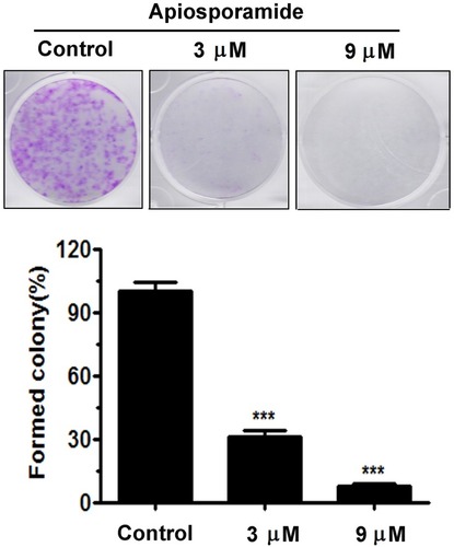
Figure 3 Apiosporamide-induced cell cycle arrest at G0/G1 phase.
Notes: (A and B) MG63 cells were treated with apiosporamide for 24 hrs. The distribution of cell cycle was assessed by flow cytometry. The percentage of cells in each phase was shown as mean± SD (n = 3). (C) MG63 cells were exposed to apiosporamide for 24 hrs. The level of cell cycle-regulated protein was analyzed by Western blotting. *P<0.05, **P<0.01 and ***P<0.001 vs vehicle control.
Abbreviation: Rb, retinoblastoma tumor suppressor protein.
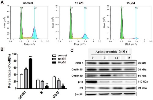
Figure 4 Apoptosis of MG63 cells following treatment with apiosporamide.
Notes: (A) SEM micrographs of apiosporamide-treated MG63 cells for 24 hrs. (B) Apoptosis was determined by an Annexin V-FITC/PI apoptosis detection kit, and representative results from flow cytometry were shown. (C) Quantitative analysis of apoptosis of MG63 cells as shown in (B). Values were presented as mean±SD (n=3). **P<0.01 and ***P<0.001 vs normal control.
Abbreviations: SEM, screening electron microscope; Annexin V-FITC, annexin V fluorescein isothiocyanate; PI, propidium iodide.
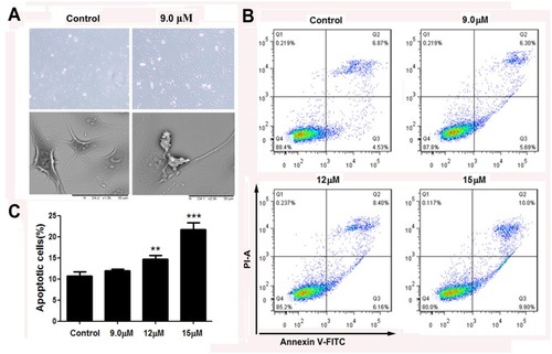
Figure 5 Effect of apiosporamide on apoptosis regulators.
Notes: (A and C) Real-time PCR analyzed the mRNA expression of caspase-3, caspase-8, caspase-9, Bax and Bcl-2 in apiosporamide-treated cells. Data were presented as mean±SD (n=3), *P<0.05, **P<0.01, ***P<0.001 compared to control cells. (B and D) MG63 cells were treated with indicated concentrations of apiosporamide for 24 hrs and protein levels of Bcl-2, Bax, caspases-3, -8 and -9 were analyzed using Western blotting.
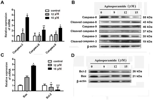
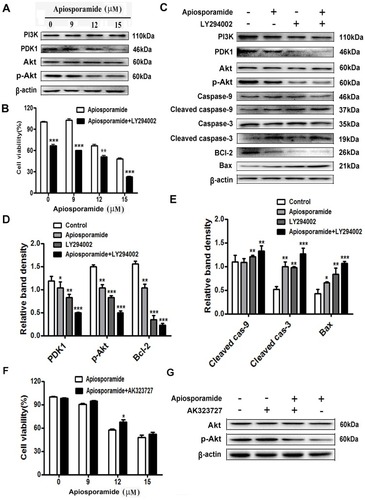
Figure 6 PI3K/Akt pathway mediated apisoporamide-induced apoptosis of MG63 cells.
Notes: (A) Cells were treated with various concentrations of apisoporamide for 24 hrs. The expressions of PI3K, PDK1, p-Akt and Akt were analyzed by Western blotting. (B) MG63 cells were incubated with LY294002 (5 μM) and apisoporamide (0, 9, 12, 15 μM) for 24 hrs. Cell viability was measured by MTT assay. Data were presented as mean±SD (n=3). (C) Western blotting results showed a significant change in the relative protein expression after MG63 cells were treated with apisoporamide (12 μM) and/or LY294002 (5 μM) for 24 hrs. (D and E) The immunoblot signals were quantified by densitometry, and mean data from independent experiments were normalized to the untreated cells. The bars are the means±SD (n = 3). (F) MG63 cells were co-treated with AK323727 (10 μM) and apisoporamide (0, 9, 12, 15 μM) for 24 hrs. Cell viability was measured by MTT assay. Data were presented as mean±SD (n=3). (G) Western blotting analysis of Akt, p-Akt and β-actin expression. MG63 cells were treated with apisoporamide (15 μM) and/or AK323727 (10 μM) for 24 hrs. *P<0.05, **P<0.01 and ***P<0.001 vs vehicle control.
Abbreviations: PI3K, phosphatidylinositol 3 kinase; PDK1, phosphoinositide-dependent kinase-1; Akt, protein kinase B.