Figures & data
Figure 1 The relationship between identified proteins and Exocarta database.
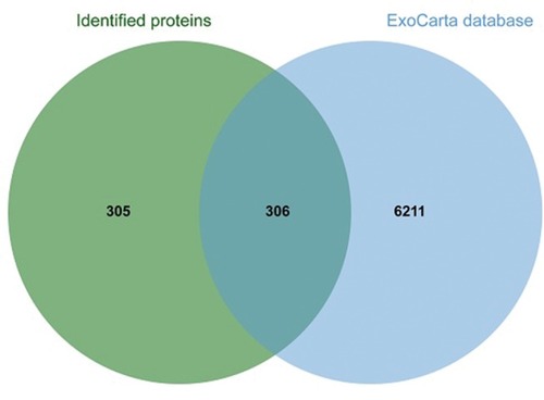
Figure 2 GO classification of proteomic data for downregulated and upregulated proteins. Differently expressed proteins were submitted to the GO classification system.
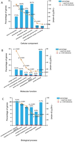
Figure 3 GO analysis shows that blood microparticle, antigen binding and receptor-mediated endocytosis were the most enriched term incellular component, molecular function and biological process.
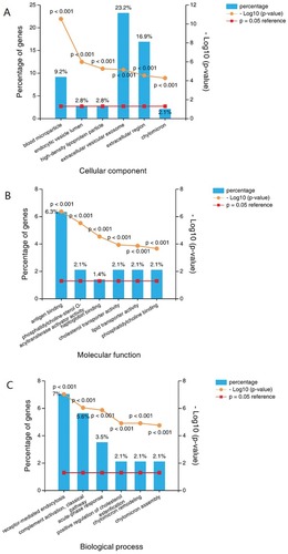
Figure 4 Network of proteins identified in overall differentially expressed proteins. Up- or downregulation is indicated by the color of nodes (upregulated in red and downregulated in green).
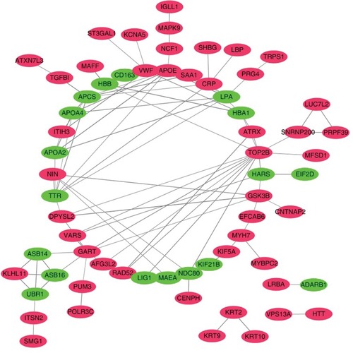
Figure 5 CCK8 test. PC cells (MiaPaCa-2, AsPC-1, 4×10⁶) were treated with serum exosomes derived from PC patients or healthy controls after 12 h of seeding (equal PDAC cells seeded first, and the OD value showed a significantly difference at 24h and 48h after treated with the exosomes, P<0.05). Compared with the control group, exosomes derived from PC patients can promote the proliferation of PC cells.
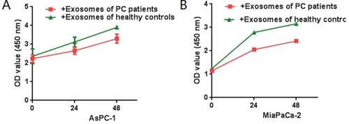
Figure 6 Wound-healing assay in vitro of PC cells (MiaPaCa-2, AsPC-1) added serum exosomes derived from PC patients. As can be seen from the figure, exosomes derived from PC patients can promote the migration of PDAC cells (***P < 0.001).
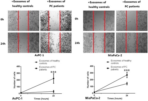
Figure 7 Transwell invasion experiment of Miapaca-2 and Aspc-1 treated with or without exosomes derived from PC patients. As can be seen from the figure, exosomes derived from PC patients can increased the trans magration of PDAC cell lines significantly (***P < 0.001).
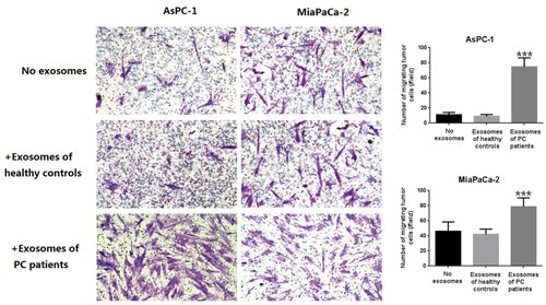
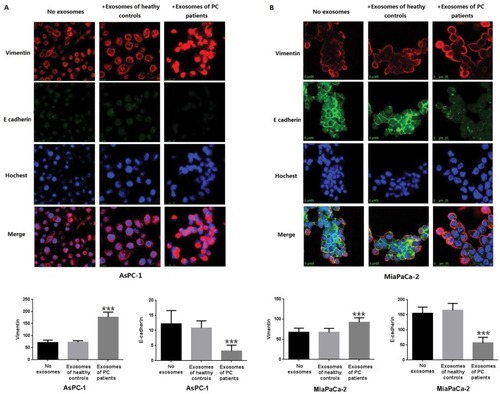
Figure 8 The laser confocal imaging of EMT related proteins. Vimentin was up expressed, while the E-cadherin was down expressed when treated with exosomes derived from PC patients. In order to make each group comparable, the fluorescence brightness of the nucleus was set at the same level in the laser confocal imaging of the three groups (***P < 0.001).