Figures & data
Table 1 Correlation Between E2F3 Expression and Clinical Characteristics of NPC Patients
Figure 1 E2F3 was upregulated in NPC cell lines and tissue samples. (A and B) E2F3 expression profiles in NPC cells and normal nasopharyngeal epithelial samples by GEO datasets GSE 12452 and GSE 53819. (C) Relative E2F3 expression in NPC tissues (n = 66) compared with that in normal tissues (n = 8). (D) E2F3 expression was detected through immunohistochemistry analysis. (E) Relationship between E2F3 expression and the clinical T stage of NPC. (F) Relationship between E2F3 expression and the lymph node metastasis of NPC. **P < 0.01.
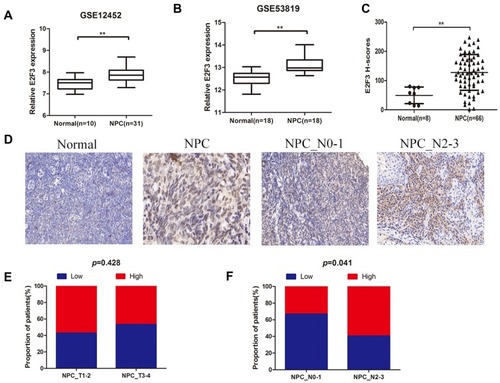
Figure 2 E2F3 promoted NPC cell invasion and migration in vitro. (A) Relative E2F3 expression in NP69 and five NPC cell lines analyzed by qRT-PCR. (B) qRT-PCR and Western blot analysis were performed to detect the efficiency of si-E2F3. (C and D) CCK8 and colony-forming assays were used to detect cell proliferation. (E) Migration of NPC cells was measured by wound-healing assay. (F) Transwell invasion assay was performed to investigate the invasive capacities of NPC cells. Data are presented as mean ± SD.**P < 0.01 compared with the control; ns, not significant.
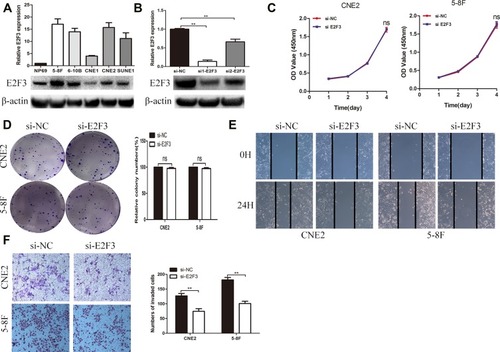
Figure 3 E2F3 acted as a target for miR-432. (A) Expression of miR-432 in NPC cells transfected. (B and C) E2F3 expression in NPC cells transfected with miR-432 mimic or inhibitor was detected through qRT-PCR. (D and E) E2F3 expression in transfected NPC cells was determined using Western blot analysis. (F) miR-432 directly bound to the 3′-UTR of E2F3. (G) Luciferase reporter assays showed that miR-432 significantly reduced the luciferase activity of the WT E2F3. **P< 0.01 compared with the control
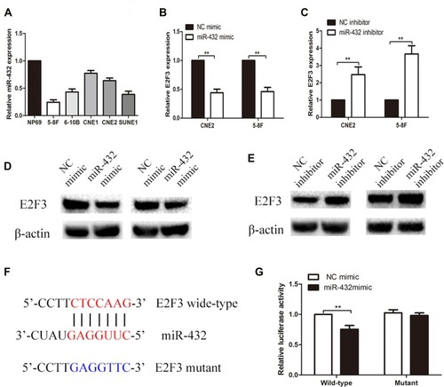
Figure 4 MiR-432 repressed the invasion and migration of NPC cells in vitro and in vivo. (A and B) Wound-healing assay and Transwell invasion assay were performed to evaluate the invasion and migration abilities of NPC cells. (C) 5-8F cells transfected with miR-432 Agomir or NC Argomir were subcutaneously injected into the plantar of each mouse (n = 5), and metastatic popliteal lymph nodes were assessed under a fluoroscope. Data are presented as mean ± SD; **P < 0.01 compared with the control.
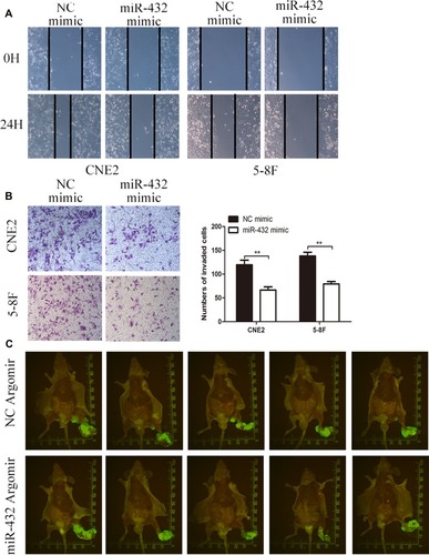
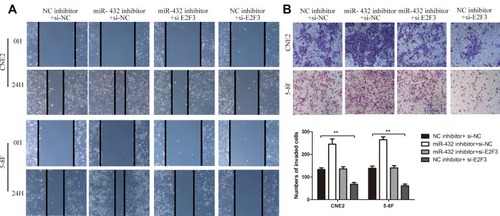
Figure 5 MiR-432 repressed the invasion and migration of NPC cells by repressing the expression of E2F3. (A) Wound-healing assay was conducted to analyze the effect of E2F3 and miR-432 on cell migration. (B) Transwell invasion assay was performed to measure the effect of E2F3 and miR-432 on cell invasion. **P < 0.01 compared with the control.