Figures & data
Figure 1 Expression pattern of circLARP4 in NSCLC clinical specimens and cells. (A) CircLARP4 expression pattern in 20 paired NSCLC tissue specimens and adjacent non-tumor tissue samples were detected by qRT-PCR. (B) CircLARP4 level in different cell lines (A549, H1299, H1975, SPC-A-1, and BEAS-2B) was examined by qRT-PCR. *P < 0.05 compared with control.
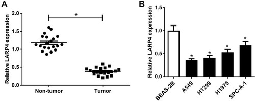
Figure 2 Effects of circLARP4 overexpression on NSCLC cell proliferation, invasion, glycolysis and apoptosis. (A) CircLARP4 expression in A549 and H1299 cells after delivery of LARP4 or pcDNA. (B) MTT assay for cell proliferation at 0 h, 24 h, 48 h, and 72 h in A549 and H1299 cells received with LARP4 or pcDNA transfection. (C) Cell invasive ability was evaluated by transwell invasion assay after A549 and H1299 cells were delivered with LARP4 or pcDNA transfection. (D) Apoptosis in A549 and H1299 cells following delivery with LARP4 or pcDNA was evaluated by flow cytometry analysis. Glucose consumption (E) and lactate production (F) in A549 and H1299 cells after introduction with LARP4 or pcDNA. (G) Western blot analysis of HK2 protein level in LARP4 or pcDNA-transfected A549 and H1299 cells. *P < 0.05 compared with negative control.
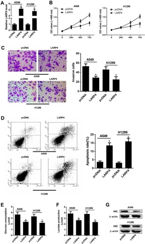
Figure 3 The association between circLARP4 and miR-135b in NSCLC cells. (A) Sequence alignment of miR-135b with binding sites in wild-type and mutant-type regions of circLARP4 was presented. The interaction between circLARP4 and miR-135b was investigated by luciferase reporter assay (B) and RIP assay (C). (D) qRT-PCR analysis of miR-135b expression in NSCLC tissues and corresponding adjacent normal tissue samples. (E) qRT-PCR analysis of miR-135b expression in different cell lines (A549, H1299, H1975, SPC-A-1, and BEAS-2B). (F and G) A549 cells were received with LARP4, si-LARP4, or respective controls transfection, followed by detection of circLARP4 and miR-135b expressions by qRT-PCR. *P < 0.05 compared with negative control.
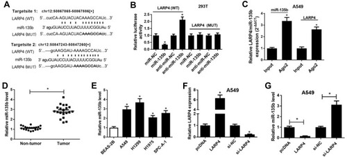
Figure 4 Effects of circLARP4 or together with miR-135b on NSCLC cell viability, glycolysis and apoptosis. A549 and H1299 cells were delivered with LARP4, pcDNA, LARP4 + miR-135b or LARP4 + miR-NC. (A) Cell viability of treated A549 and H1299 cells was detected by MTT assay. (B) Cell invasion potential was assessed by transwell invasion assay. (C) The percentage of apoptotic cells in the transfected A549 and H1299 cells was detected by flow cytometry analysis. (D and E) Glucose consumption and lactate production in the introduced A549 and H1299 cells. (F) HK2 protein level in the transfected A549 and H1299 cells was measured by Western blot. *P < 0.05 compared with negative control.
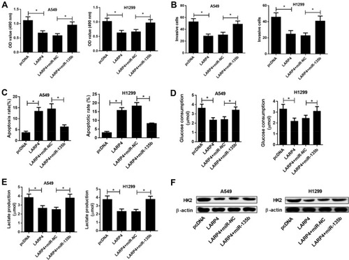
Figure 5 Effects of circLARP4 on NSCLC tumor growth and miR-135b expression in vivo. (A and B) CircLARP4 and miR-135b expressions in the xenograft tumors in nude mice derived from subcutaneous injection of A549 cells transfecting with lenti-LARP4 or lenti-con were estimated by qRT-PCR. (C) After 5 weeks of implantation, the xenograft tumors were dissected and weighed. (D) Tumor formation was monitored weekly for 5 weeks. *P < 0.05 compared with negative control.
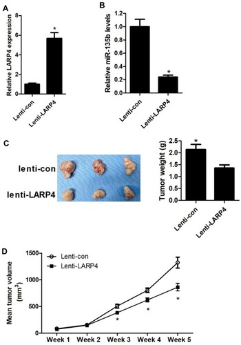
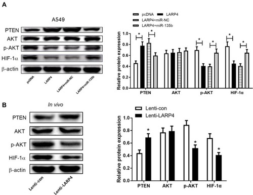
Figure 6 Effects of circLARP4 or along with miR-135b on the PTEN/AKT/HIF-1α signaling pathway in NSCLC. (A) The protein levels of PTEN, p-AKT, AKT and HIF-1α in A549 cells transfected with LARP4, pcDNA, LARP4 + miR-135b or LARP4 + miR-NC were detected by Western blot analysis. (B) Western blot analysis of PTEN, p-AKT, AKT and HIF-1α protein levels in xenograft tumor tissues. *P < 0.05.