Figures & data
Table 1 Numbers of Specimens for Three Detection Methods
Table 2 The Expression of α-Subunit in Normal Laryngeal Tissues in Three Detection Methods
Figure 1 Expression levels of the α- and β-subunits of H+/K+-ATPases (proton pumps) in normal laryngeal tissues as revealed by immunohistochemical staining. (A) Gastric control tissues stained strongly for the α-subunit. (B) Gastric control tissues stained strongly for the α-subunit. (C) The α-subunit was expressed in normal epiglottic mucosa. (D) The α-subunit was expressed in normal epiglottic mucosa. (E) The α-subunit was expressed in normal false vocal cord mucosa. (F) The α-subunit was expressed in normal false vocal cord mucosa. (G) The α-subunit was expressed in normal epiglottic cartilage. (H) The α-subunit was expressed in normal epiglottic cartilage.
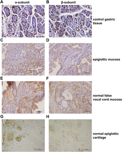
Table 3 The Expression of β-Subunit in Normal Laryngeal Tissues in Three Detection Methods
Figure 2 Expression levels of mRNAs encoding the H+/K+-ATPase α- and β-subunits in normal laryngeal tissues and laryngeal carcinomas as revealed by real-time RT-PCR. (A) α-subunit mRNA, (B) β-subunit mRNA. *P < 0.05; **P < 0.01.
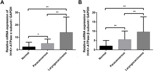
Figure 3 Expression levels of the H+/K+-ATPase α- and β-subunit proteins in representative normal laryngeal tissues and laryngeal carcinomas as revealed by Western blotting. (A) α-subunit protein, (B) β-subunit protein.
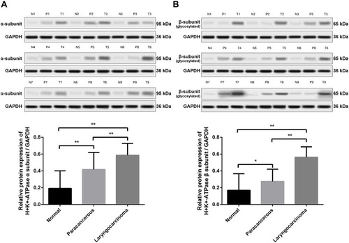
Table 4 The Relationship Between the Expression of α-Subunit or β-Subunits and Clinicopathological Features of Laryngeal Carcinoma
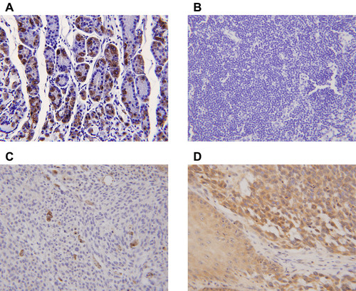
Figure 4 Expression of the α-subunit of H+/K+-ATPases (proton pumps) in laryngeal carcinomas as revealed by immunohistochemical staining. (A) Gastric control tissues exhibited strong staining for the α-subunit. (B) The α-subunit was not expressed in paracarcinoma tissues. (C) The α-subunit was not expressed in laryngeal carcinomas. (D) Positive staining (brown) is evident in both the cytoplasm and plasma membrane of laryngeal carcinoma cells.
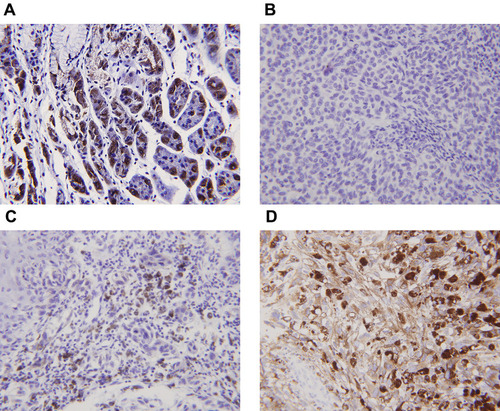
Figure 5 Expression of the β-subunit of H+/K+-ATPases (proton pumps) in laryngeal carcinomas as revealed by immunohistochemical staining. (A) Gastric control tissues exhibited strong staining for the β-subunit. (B). The β-subunit was not expressed in paracarcinoma tissues. (C) The β-subunit was not expressed in laryngeal carcinomas. (D) Positive staining (brown) is evident in both the cytoplasm and plasma membrane of laryngeal carcinoma cells.