Figures & data
Figure 1 The pathologic diagnosis of an endoscopic biopsy of rectal.
Notes: Neoplasm composed of pleomorphic epithelial cells arranged in glandular pattern (hematoxylin and eosin staining, ×40) (A); Immunohistochemical exam positive for CDX2 (B).
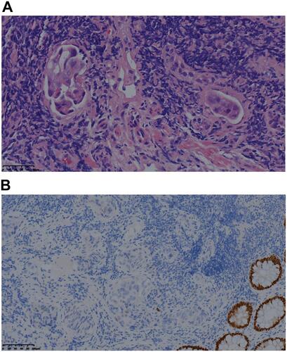
Figure 2 The evolution of skin metastases.
Notes: In September 2019, the skin of the hypogastrium and perineum was red and swollen with a rubbery appearance, a rash-like swelling on the surface, and local fusion (A); In December 2019, the skin nodules had increased in size and involved the skin of the hypogastrium, left thigh, bilateral groin, and perineum. These nodes mixed together, and formed tiny open sores, or ulcers, on the surface of the nodes (B); After palliative treatment with FOLFIRI plus cetuximab and vemurafenib, the cutaneous nodules decreased in size (C).
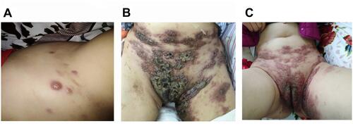
Figure 3 Pathological findings of rectal carcinoma skin metastases.
Notes: The pathologic diagnosis of skin nodule was metastatic rectal adenocarcinoma (hematoxylin and eosin staining, ×40) (A); Negative CDX2 staining in presence of skin metastases of rectal adenocarcinoma (DAB, ×10) (B); Positive SATB2 staining in presence of skin metastases of rectal adenocarcinoma (DAB, ×20) (C); Negative CK7 staining in presence of skin metastases of rectal adenocarcinoma (DAB, ×20) (D).
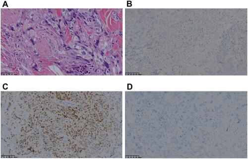
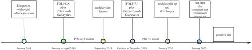
Figure 4 Timeline of diagnosis and treatment of the patient.