Figures & data
Figure 1 The main regulatory pathway of PD-L1 in cancer in KEGG database.
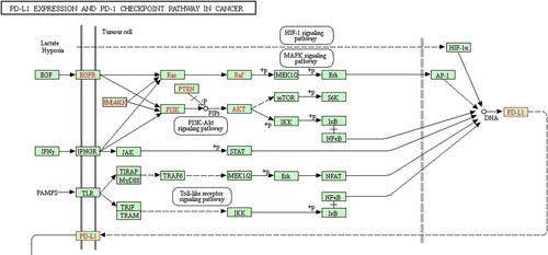
Table 1 The Baseline Characteristics of Gastric Cancer Patients
Figure 2 The expression of PD-L1-related regulatory factors is abnormal in GC patients. (A) IHC detected the expression of PD-L1, HIF-1, EGFR, IFNGR and TLR4. (B) ELISA detected the expressions of EGF, IFNG and IL-6 in serum of patients. ***P<0.001 vs normal; ##P<0.01, ###P<0.001 vs patient.
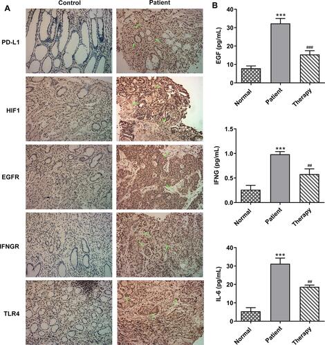
Table 2 The Targeting Effect of BXXX and Its Main Components on Molecules
Figure 3 BXXX and its major components target and regulate key factors in the PD-L1 pathway.
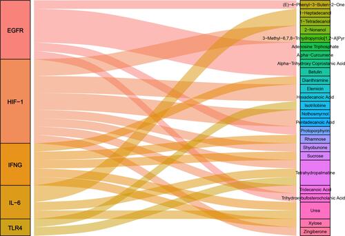
Figure 4 BXXX inhibits the proliferation and promotes the apoptosis of GC cells by indirectly regulating the expression of PD-L1 through multiple pathways and targets. (A) CCK-8 detected the cell viability. (B) Tunel assay detected the apoptosis of cells. (C) Western blot detected the expression of related proteins in the PD-L1 pathway. (D) Western blot detected the expression of EGFR, IFNGR and TLR4 in cell membrane. *P<0.05, **P<0.01, ***P<0.001 vs NC. Control group: cells were cultured in normal medium; NC group: cells were cultured in BXXX-free rat serum; BXXX-5%: cells were cultured in BXXX-5% rat serum; BXXX-10%: cells were cultured in BXXX-10% rat serum; BXXX-20%: cells were cultured in BXXX-20% rat serum.
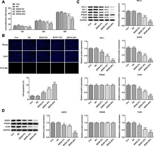
Figure 5 It was further found that BXXX inhibits the proliferation and promotes the apoptosis of GC cells by indirectly regulating the expression of PD-L1 through multiple pathways and targets. (A) CCK-8 detected the cell viability. (B) Tunel assay detected the apoptosis of cells. (C) Western blot detected the expression of related proteins in the PD-L1 pathway. (D) Western blot detected the expression of EGFR, IFNGR and TLR4 in cell membrane. *P<0.05 vs control; ***P<0.001 vs Con; #P<0.05, ##P<0.01, ###P<0.001 vs BXXX.
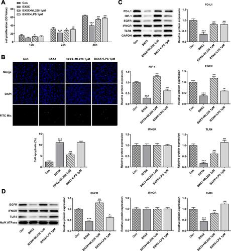
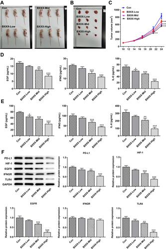
Figure 6 BXXX inhibits tumor formation in GC-bearing mice via multiple targets and pathways. (A) Pictures of each group of mice. (B) Pictures of tumor tissues of mice in each group. (C) Statistical analysis diagram of tumor tissue volume. ELISA detected the expressions of EGF, IFNG and IL-6 in animal tissues (D) and serum (E). (F) Western blot detected the expressions of related proteins in the PD-L1 pathway. *P<0.05, **P<0.01, ***P<0.001 vs Con.