Figures & data
Figure 1 ACTL8 expression was upregulated in TNBC and associated with the poor prognosis of TNBC. (A) TCGA dataset analysis showing the expression of ACTL8 in different molecular subtypes of breast cancer. (B) ACTL8 protein expression in TNBC cells was determined by Western blot. (C) mRNA expression of ACTL8 in TNBC cells was determined by qRT-PCR. (D) Kaplan–Meier survival analysis of overall survival based on TCGA data. **P < 0.01, vs normal group (A); **P < 0.01, vs MCF-10A cells group (B and C).
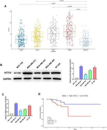
Figure 2 Silencing ACTL8 suppressed the proliferation in MDA-MB-231 and BT-549 cells. (A) ACTL8 protein expression in TNBC cells transfected with control, si-NC orsi-ACTL8 was determined by Western blot. (B) ACTL8 mRNA expression in TNBC cells transfected with control, si-NC orsi-ACTL8 was determined by qRT-PCR. (C) EdU assay was used to assess the proliferation of the transfected MDA-MB-231 and BT-549 cells. (D) Colony formation assay was executed to detect the colony numbers of the transfected MDA-MB-231 and BT-549 cells. **P < 0.01, vs Control and si-NC group.
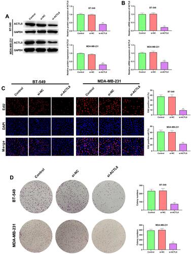
Figure 3 Silencing ACTL8 facilitated the apoptosis in MDA-MB-231 and BT-549 cells. (A) Apoptosis was assessed in transfected MDA-MB-231 and BT-549 cells by flow cytometry. (B) The expression of Bcl-2, Bax and cleaved caspase-3 in transfected MDA-MB-231 and BT-549 cells was measured by Western blot. **P < 0.01, vs Control and si-NC group.
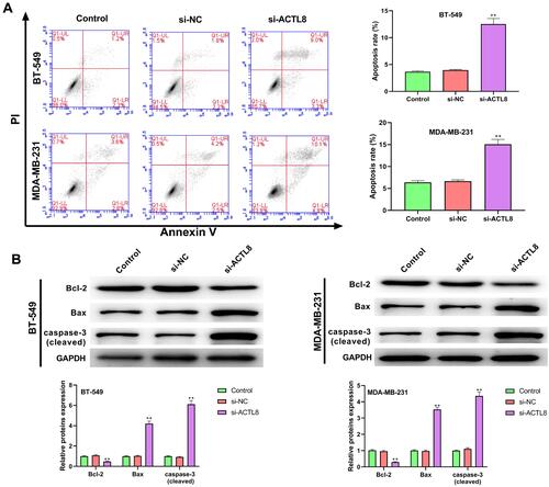
Figure 4 Silencing ACTL8 suppressed the migration and invasion abilities in MDA-MB-231 and BT-549 cells. (A) Wound healing assay was used to assess the migration of the transfected MDA-MB-231 and BT-549 cells. (B) Transwell assay was executed to detect the migration of the transfected MDA-MB-231 and BT-549 cells. (C) Transwell assay was executed to detect the invasion of the transfected MDA-MB-231 and BT-549 cells. **P < 0.01, vs Control and si-NC group.
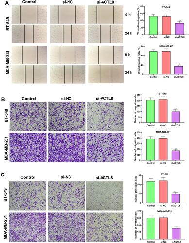
Figure 5 Silencing ACTL8 inhibited the activation of PI3K/AKT/mTOR signaling pathway in MDA-MB-231 and BT-549 cells. (A) The expression of p-PI3K, PI3K, p-AKT, AKT, p-mTOR and mTOR in transfected BT-549 cells was measured by Western blot. (B) The expression of p-PI3K, PI3K, p-AKT, AKT, p-mTOR and mTOR in transfected MDA-MB-231cells was measured by Western blot. **P < 0.01, vs Control and si-NC group.
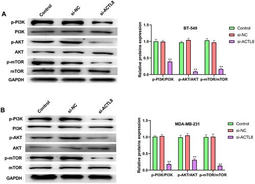
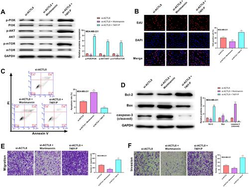
Figure 6 PI3K/AKT/mTOR signaling pathway was involved in ACTL8 modulated the proliferation, apoptosis, migration and invasion in MDA-MB-231 cells. (A) After treatment of PI3K/AKT/mTOR pathway inhibitor (Wortmannin) and PI3K/AKT/mTOR pathway activator (740Y-P), the expression of p-PI3K, PI3K, p-AKT, AKT, p-mTOR and mTOR in transfected MDA-MB-231cells was measured by Western blot. (B) EdU assay was used to assess the proliferation of the transfected MDA-MB-231 cells following the treatment of Wortmannin and 740Y-P. (C) Flow cytometry was performed to assess the apoptosis of the transfected MDA-MB-231 cells following the treatment of Wortmannin and 740Y-P. (D) After treatment of Wortmannin and 740Y-P, the expression of Bcl-2, Bax and cleaved caspase-3 in transfected MDA-MB-231cells was measured by Western blot. (E) Transwell assay was used to assess the migration of the transfected MDA-MB-231 cells following the treatment of Wortmannin and 740Y-P. (F) Transwell assay was used to assess the invasion of the transfected MDA-MB-231 cells following the treatment of Wortmannin and 740Y-P. **P < 0.01, vs si-ACTL8 group.