Figures & data
Figure 1 (A) NGS confirmed ALK fusion (EML4 exon 13–ALK exon 20, variant allele frequency was 4192); (B) EGFR 18 exon (c.2156G>C:55241708, p. G719A, abundance 74.8%); (C) EGFR exon 19 (c2239T>G:55242469, pL747V, abundance 70.05%).
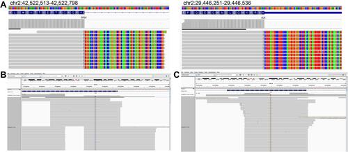
Figure 2 (A and B) Thoracic CT scan detected a subpleural soft tissue mass in the right upper lung (red arrows); (C and D) after 4 weeks of treatment with afatinib, a thoracic CT scan showed a diminished subpleural tumor in the right upper lung.
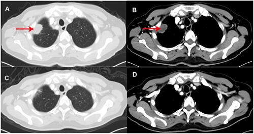
Figure 3 (A) Brain MRI detected abnormal enhancement in the brain (red arrows) and (B and C) leptomeningeal linear enhancement (red arrows) in the cerebellar hemisphere; (D) brain MRI showed that the abnormal enhancement in the brain and (E and F) the leptomeningeal linear enhancement in the cerebellar hemisphere had disappeared.
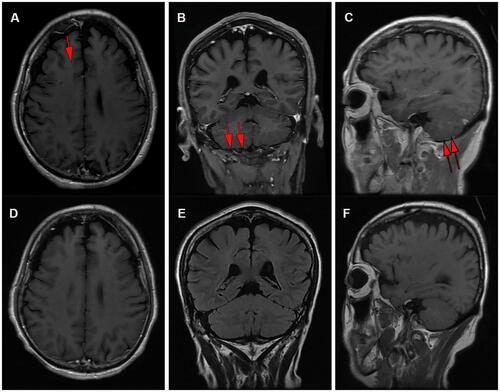
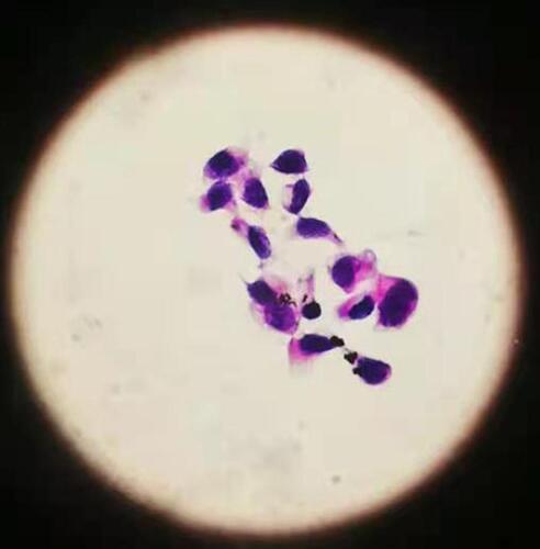
Figure 4 Lumbar puncture indicated positive cytology of cerebrospinal fluid (at high magnification 10 * 40).