Figures & data
Figure 1 Immunohistochemical staining of angioimmunoblastic T-cell lymphoma tissue sample. Immunohistochemical staining of H&E (A) and Ki67 (I), negative for CD20 (B) and positive for CD3 (C), CD21 (D), CD10 (E), Bcl-6 (F), CD4 (G), PD-1 (H) (A: H&E×100, inset: H&E×400, B–I: ×200).
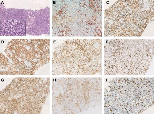
Figure 2 Immunohistochemical staining of diffuse large B-cell lymphoma tissue sample. Immunohistochemical staining of H&E (A) and Ki67 (I), positive for CD20 (B), MUM1 (D), CD10 (E), Bcl-6 (F), EBER (G) and negative for CD3 (C), PD-1 (H) (A: H&E×100, inset: H&E×400, B–H: ×200, I: ×100).
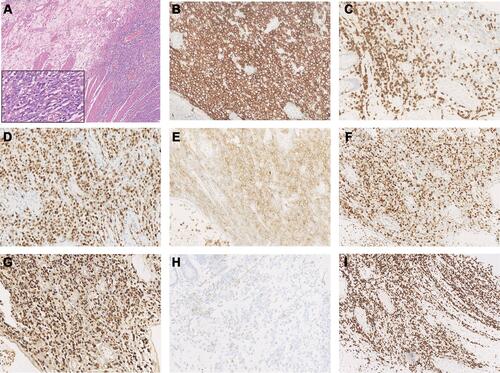
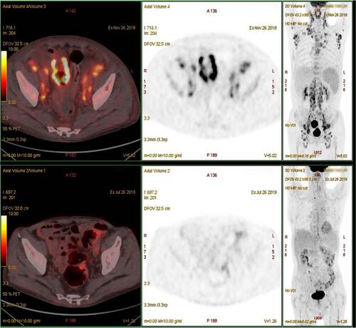
Figure 3 Comparison of positron emission tomography/computed tomography scans before and after lenalidomide-combination treatment.