Figures & data
Table 1 The Primers for Real-Time PCR and Quantitative RT-PCR That Were Used in This Study
Figure 1 CDH11 is downregulated in a highly metastatic tongue squamous cell carcinoma cell line. (A) Quantitative real-time PCR was performed on LN4 and TSCC cell line CAL27 to explore the expression of the cadherin family (ND means not detectable). (B) 44 normal samples and 502 head and neck squamous cell carcinoma samples from the TCGA database. (C) 15 normal samples and 147 TSCC samples from the TCGA database. (D) Clinical samples from 57 cases of cancer and adjacent normal tissues were evaluated with respect to expression by the 2−ΔΔCt method. P>0.05.
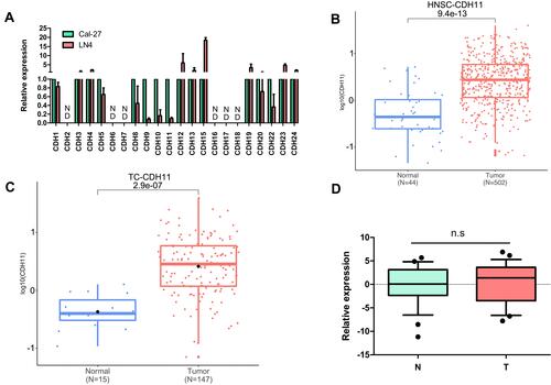
Figure 2 Overexpression of CDH11 in TSCC cells. The expression of CDH11 were confirmed by real-time PCR (A and B) and Western blotting (C and D) in the TSCC cells after infected with lentivirus and selected with puromycin. (E) Cellular immunofluorescence experiments confirmed the CDH11 expression quantities presented on the membranes of the experimental group. (400X, *P < 0.05, **P < 0.01).
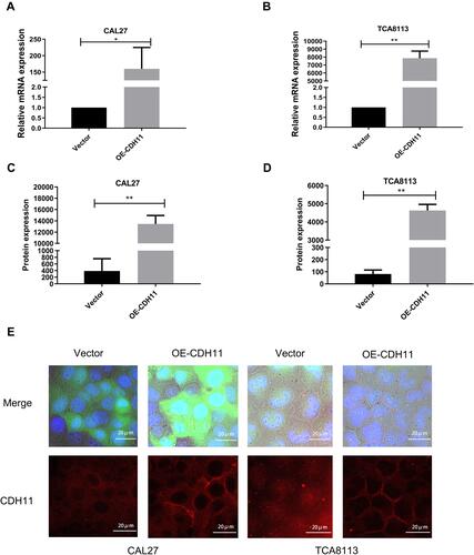
Figure 3 Overexpression of CDH11 did not affect the proliferation and stemness of TSCC cells. The proliferation ability of TSCC cells was measured by the CCK-8 assay (A) and colony formation assay (B) after upregulating CDH11.(C) The cancer stem cell sphere formation assay was adopted to test the stemness of tongue cancer cells. (n.s, nonsignificance, P>0.05).
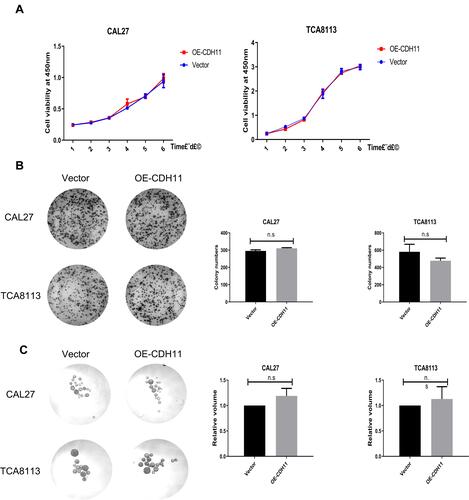
Figure 4 Overexpression of CDH11 did not affect the migration and invasion of TSCC cells. (A) Representative images for migration assay (upper panel) or invasion assay (lower panel) of the CAL27 and TCA8113 cells after infection with CDH11 lentivirus. (B and C) The numbers of cells crossing the transwell chambers were counted and analyzed. (n.s, nonsignificance, P>0.05).
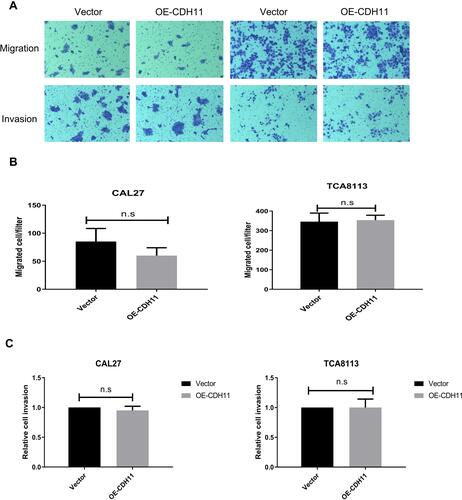
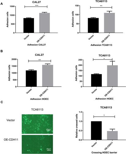
Figure 5 Overexpression of CDH11 inhibits TSCC cell metastatic potential in vitro (A) in the cell adhesion assay, we inoculated the cells upon a monolayer of corresponding homogeneous cells to test its adhesion ability. (B) Like before, we inoculated the cells upon a monolayer of HOECs to test its adhesion ability. (C) We tested their ability to pass through the single layer of HOECs in the transcellular migration assay. (100X, *P < 0.05, **P < 0.01, ***P < 0.001).