
Figures & data
Figure 1 Primary signet-ring cell carcinoma of the uterine cervix in the biopsy. (A) Colposcope image of the cervical mass. (B) Histopathological findings of the cervical (left) and ovarian (right) lesion (hematoxylin and eosin stain, ×200 magnification).
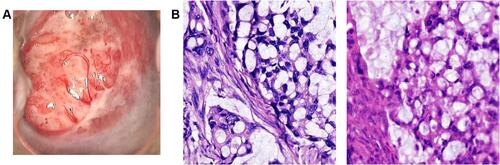
Figure 2 Ovarian involvement associated with the cervical adenocarcinomatous lesion. (A) Computed tomography depicting the pelvic lesion with a solid-cystic appearance. (B) The cervix and right ovary during surgery.
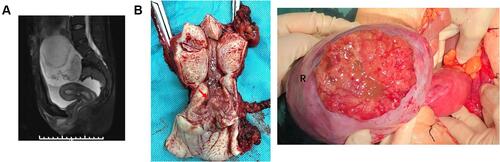
Figure 3 Immunohistochemical staining and polymerase chain reaction results of the cervical lesion. (A) Tumor cell nuclei were negative for estrogen receptor. (B) The presence of human type 18 papillomavirus in cervical and ovarian tissue.
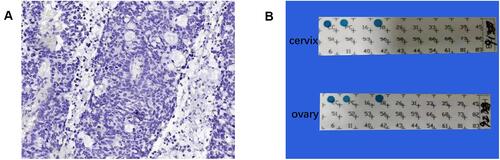
Table 1 Summary of Cases of Primary Signet-Ring Cell Carcinoma of the Cervix, Modified from Giordano, Sal, Hamada et al
Table 2 Key Points of Primary Signet-Ring Cell Carcinoma of the Uterine Cervix (PSRCCC) with Ovarian Involvement