Figures & data
Figure 1 β-Elemene enhanced the cytotoxic effect of Dox in Dox-resistant osteosarcoma cells. (A) MG63 and MG63/Dox cells or (B) Saos-2 and Saos-2/Dox cells were treated with 0, 0.5, 2, 5, 25 or 50 μg/mL Dox for 48 h respectively. CCK-8 assay was used to detect cell viability. **P < 0.01, compared with the MG63 or Saos-2 group. (C) MG63 and Saos-2 cells were treated with 0, 5, 10, 20, 50 or 100 μg/mL β-Elemene for 48 h respectively. CCK-8 assay was used to detect cell viability. *P < 0.05, **P < 0.01, compared with the β-Elemene (0 μg/mL) group. (D) MG63/Dox and Saos-2/Dox cells were treated with 0, 25, 50, 100 or 200 μg/mL β-Elemene for 48 h respectively. CCK-8 assay was used to detect cell viability. *P < 0.05, **P < 0.01, compared with the β-Elemene (0 μg/mL) group. (E) MG63/Dox cells or (F) Saos-2/Dox cells were treated with Dox (0, 0.5, 2, 5, 25 or 50 μg/mL), or Dox (0, 0.5, 2, 5, 25 or 50 μg/mL) plus 25 μg/mL β-Elemene for 48 h. CCK-8 assay was used to detect cell viability. *P < 0.05, **P < 0.01, compared with the Dox treatment group.
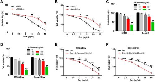
Table 1 Evaluation of Combination of Dox with β-Elemene in MG63/Dox and Saos-2/Dox Cells (48 h Treatment)
Figure 2 Combination of Dox with β-Elemene inhibited the proliferation of Dox-resistant osteosarcoma cells. (A) MG63/Dox cells or (B) Saos-2/Dox cells were treated with 5 μg/mL Dox or/and 25 μg/mL β-Elemene for 48 h. EdU staining assay was used to determine cell proliferation. *P < 0.05, **P < 0.01, compared with the control group; ##P < 0.01, compared with the Dox treatment group.
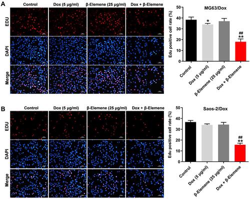
Figure 3 Combination of Dox with β-Elemene induced the apoptosis of Dox-resistant osteosarcoma cells. MG63/Dox and Saos-2/Dox cells were treated with 5 μg/mL Dox or/and 25 μg/mL β-Elemene for 48 h. (A and B) Apoptotic cells were measured by flow cytometry. (C) Expression levels of Bcl-2, Bax and cleaved caspase 3 in cells were detected with Western blotting. β-actin was used as an internal control. (D–F) The relative expressions of Bcl-2, Bax and cleaved caspase 3 in cells were quantified via normalization to β-actin. *P < 0.05, **P < 0.01, compared with the control group; ##P < 0.01, compared with the Dox treatment group.
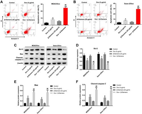
Figure 4 Combination of Dox with β-Elemene induced the oxidative stress in Dox-resistant osteosarcoma cells. MG63/Dox and Saos-2/Dox cells were treated with 5 μg/mL Dox or/and 25 μg/mL β-Elemene for 48 h. (A and B) Flow cytometry was applied to assess the ROS production in MG63/Dox and Saos-2/Dox cells. (C and D) ELISA assay was used to detect the level of GSH in the supernatants of MG63/Dox and Saos-2/Dox cells. *P < 0.05, **P < 0.01, compared with the control group; #P < 0.05, ##P < 0.01, compared with the Dox treatment group.
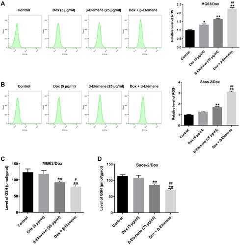
Figure 5 β-Elemene enhanced the sensitivity of Saos-2/Dox cells to Dox via downregulation of Prx-1. (A) Western blot assay was used to detect the expression of Prx-1 in MG63 and MG63/Dox cells. β-actin was used as an internal control. **P < 0.01, compared with the MG63 group. (B) Western blot assay was used to detect the expression of Prx-1 in Saos-2 and Saos-2/Dox cells. β-actin was used as an internal control. **P < 0.01, compared with the Saos-2 group. (C) MG63/Dox and Saos-2/Dox cells were treated with 5 μg/mL Dox or/and 25 μg/mL β-Elemene for 48 h. The expression level of Prx-1 in MG63/Dox and Saos-2/Dox cells was detected with Western blotting. (D) The relative expression of Prx-1 in cells was quantified via normalization to β-actin. **P < 0.01, compared with the control group; ##P < 0.01, compared with the Dox treatment group. (E) Western blot analysis of Prx-1 expression in MG63/Dox and Saos-2/Dox cells transfected with pcDNA3.1-Prx-1. **P < 0.01, compared with the pcDNA3.1-ctrl group. (F) MG63/Dox and Saos-2/Dox were treated with 5 μg/mL Dox and 25 μg/mL β-Elemene, or the combination of Dox and β-Elemene and pcDNA3.1-Prx-1 for 48 h. CCK-8 assay was used to detect cell viability. (G) Western blot analysis of Prx-1 expression in MG63/Dox and Saos-2/Dox cells. **P < 0.01, compared with the control group; ##P < 0.01, compared with the Dox + β-Elemene treatment group.
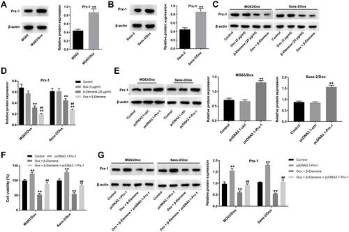
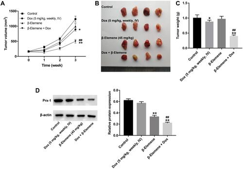
Figure 6 Combination of Dox with β-Elemene inhibited tumorigenesis in Saos-2/Dox xenograft in vivo. (A) Tumor volumes of mice were monitored at different time points. (B and C) Saos-2/Dox xenograft tumors in each group was pictured and weighted on day 21. (D) Western blot analysis of Prx-1 expression in tumor tissues. *P < 0.05, **P < 0.01, compared with the control group; ##P < 0.01, compared with the Dox treatment group.