Figures & data
Figure 1 A large tumor in patient’s right breast upon first clinical examination. The tumor had an exogenous growth with an ulcer measuring 5×5 cm in the center, covered with white and yellow exudate.
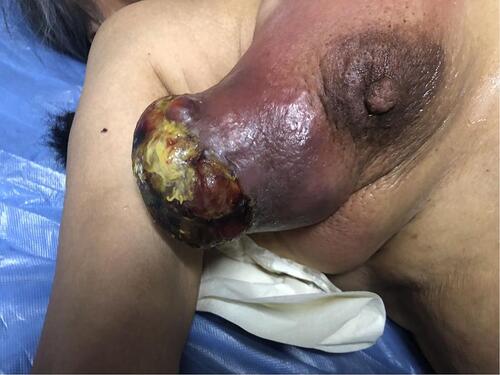
Figure 2 The outcome of breast and axillary B ultrasonography. (A) B ultrasonography showed a 12.5-cm diameter mass in the lateral quadrant of the right breast. (B) B ultrasonography showed enlarged axillary lymph nodes. The larger node had a diameter of 1.2 cm.
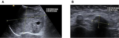
Figure 3 Results of chest computed tomography (CT). (A) The red arrow shows a large breast tumor with thickened skin. (B) The red arrow shows a nodule in the upper lobe of the right lung, indicating lung metastasis. (C) The red arrow shows another nodule in the upper lobe of the right lung, suggesting lung metastasis. (D) The red arrow shows abnormal bone density on the thoracic third vertebral body (T3), indicating bone metastasis.
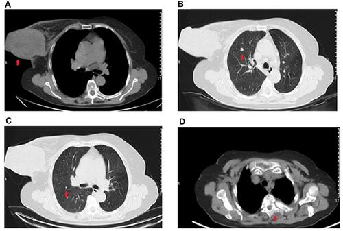
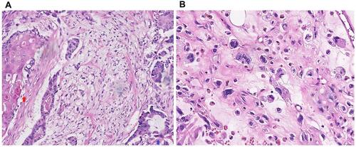
Figure 4 Results of pathological examination. (A) Blue arrow shows invasive ductal carcinoma, while the red arrow shows squamous cell carcinoma. (B) High-grade sarcoma.