Figures & data
Figure 1 Experimental study design.
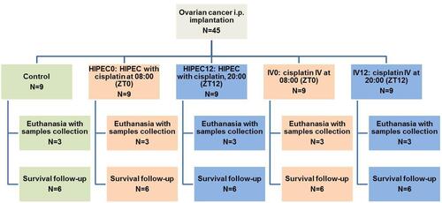
Figure 2 Daily variation in proliferation in small intestine crypts, (B) apoptosis in small intestine crypts, and (C) DNA damage/repair in liver in a pilot study. (D) Representative photomicrograph of anti-γ-H2AX stained liver of tumor-bearing rat (×400), ZT6. Control: intact rats; OC: rats with ovarian cancer. **–p<0.01, ***–p<0.001, ****p<0.0001 compared to control at the same time point, #p<0.05 compared to ZT0, ####p<0.0001 compared to ZT0.
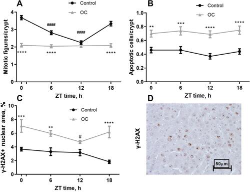
Figure 3 Effects of HIPEC and IV cisplatin administration at ZT0 and ZT12 on (A) index of mitotic activity in tumor, (B) DNA damage/repair in liver (γ-H2AX-immunostaining, (C) mitotic activity, and (D)apoptosis in intestinal epithelium (mean ± SEM). *p<0.05, **p<0.01, ***p<0.001, ****p<0.0001 compared to control, ap<0.05, aap<0.01, aaa p<0.001, aaaap<0.0001 compared to different treatment at the same time, #p<0.05, ##p<0.01, ###p<0.001, ####p<0.0001 compared to the same treatment at the different time (Tukey’s test).
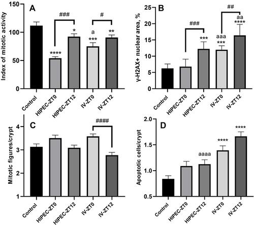
Figure 4 Representative microphotographs of anti-γ-H2AX-stained kidney sections, illustrating samples from: (A) group receiving HIPEC with cisplatin at 08:00 (ZT0), (B) group receiving IV cisplatin at 08:00 (ZT0), (C) group receiving HIPEC with cisplatin at 20:00 (ZT12), (D) group receiving IV cisplatin at 20:00 (ZT12). Black arrows: positively stained nuclei; red arrows: cystic tubular changes (×200).
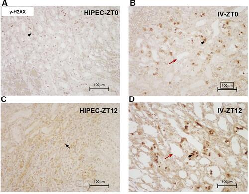
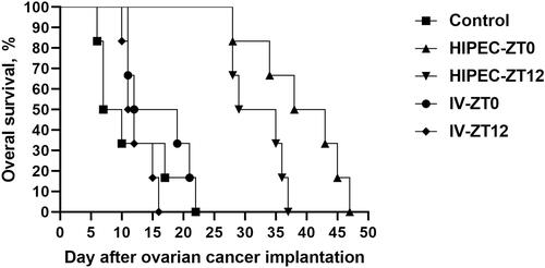
Figure 5 Survival of rats with ovarian cancer after intravenous administration and HIPEC with cisplatin at different time points.