Figures & data
Figure 1 Chest computed tomography (CT) findings. (A) Chest CT showing a round nodule-like shadow in the left upper lobe, with a clear boundary and shallow lobule of about 2.5 cm diameter. (B) After anti-infection treatment, the chest CT showed a high-density shadow in the tongue segment of the left lung, with a size of about 3.2 cm * 2.7 cm, likely to be neoplastic. (C) Chest CT showing a 5.3 cm * 3.5 cm tumor mass in the lingual segment of the left upper lobe. (D) The lesion in the left upper lobe was about 9.1 cm * 6.7 cm, with bubbles and liquid dark areas. After enhanced CT, the lesion showed circular enhancement. (E) Chest CT showing a mass of 8.8 cm * 8.0 cm in the upper lobe of the left lung, containing gas-liquid plane. (F) Chest CT showing a small nodular high-density shadow in the lower lobe of right lung with clear boundary and 1.2 cm diameter.
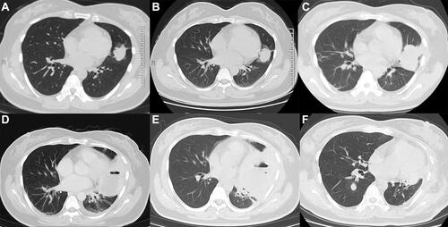
Figure 2 Bronchoscopy revealing congestion and edema of the mucosa at the mouth of the left lingual duct.
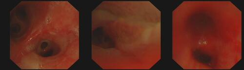
Figure 3 Hematoxylin and eosin staining of a tumor section (×200). (A) Pathological examination under light microscopy, showing a poorly differentiated non-small cell carcinoma occupying the space of the upper lobe of the left lung. (B) Pathological examination under light microscopy, showing a poorly differentiated carcinoma of the right breast with extensive hemorrhagic necrosis. (C) Immunohistochemical staining for β-hCG (positive in foci).
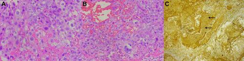
Figure 4 Breast ultrasound showing hypoechoic mass in the right breast (BI-RADS 4a) (green box).
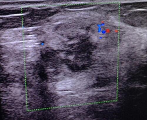
Figure 5 Chest computed tomography (CT) before and after chemotherapy. (A) Chest CT showing multiple nodules and masses in both lungs, the larger of which was about 5.7 cm * 3.4 cm. (B) Chest CT after chemotherapy showing tumors smaller than those before.
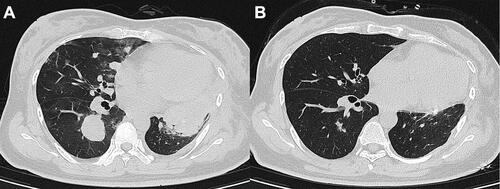
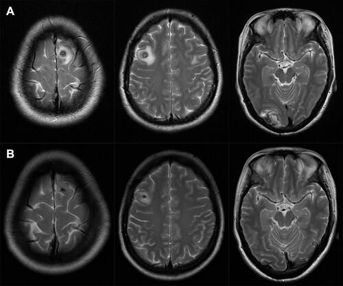
Figure 6 Brain magnetic resonance imaging (MRI) before and after chemotherapy. (A) Brain MRI before chemotherapy shows multiple nodules in bilateral frontal lobe and right occipital lobe, suggesting metastasis with a little bleeding. (B) After chemotherapy, brain MRI shows that bilateral frontal lobe and right occipital lobe nodules were smaller than before, and the peripheral edema was relieved.