
Figures & data
Figure 1 Colposcopes view of the lesion. (A) An 8-millimeter-diameter intravascular papillary endothelial hyperplasia in the right apical angle of the vagina with medium amount of blood. The lesion is red to purple coloration, and a medium amount of blood was seen. (B) Iodine staining of the lesion.
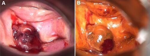
Figure 2 Pathology and immunohistochemistry result (200×). (A) Microscopic finding presents proliferation of erythrocytes within a dilated vascular structure. (B) Ki-67 labeling index was about 5%. Immunohistochemistry shows the tissue was positive for CD31 (C) and CD34 (D).
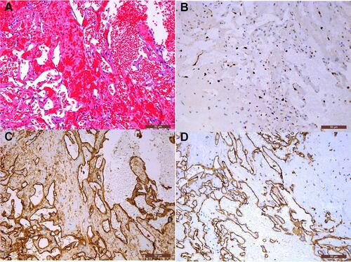
Table 1 Summary of Reports of Masson’s Tumor Occurred in the Female Reproductive System