
Figures & data
Figure 1 (A) Chest CT detected a 13×4 mm nodule in the right upper lung; (B) abdominal MRI revealed a perirenal tumor, 18×28 mm in size, close to the hilum of the left kidney.
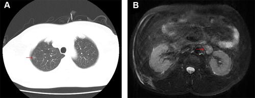
Figure 2 Positron emission tomography (PET)-CT whole body imaging. (A) A 15×10 mm shadow with ground glass density in the upper right lung was detected; (B) a neoplastic lesion with hypermetabolism close to the left renal hilum was shown.
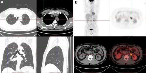
Figure 3 Immunohistochemistry of the perirenal mass showed positive for Melan-A, cluster differentiation (CD)56, and TFE3, while negative for HMB45, Syn, S-100 and SOX10. Ki67 was present in approximately 1% of tumor cells.
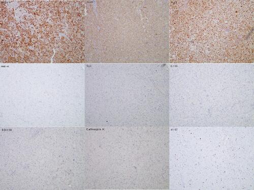
Table 1 Characteristics of Prior Cases of Perirenal Perivascular Epithelioid Cell Tumor (PEComa)