Figures & data
Table 1 Distribution of AATF in Bladder Cancer According to Clinicopathological Characteristics
Figure 1 Expression of AATF in bladder cancers. (A) Weak AATF staining in normal bladder uroepithelial tissue. (B) Negative AATF staining in a bladder cancer. (C) Moderate AATF staining in a case of bladder cancer. (D) Strong cytoplasmic and nuclear AATF staining in a case of bladder cancer. (E) Analysis of Dyrskjot bladder 3 Oncomine dataset suggested that AATF mRNA was increased in bladder cancers compared with normal bladder tissues. (F) Analysis of Lee bladder dataset suggested that AATF mRNA was higher in bladder cancer tissues compared with normal bladder tissues. (G) Analysis of Sanchez-Carbayo bladder 2 suggested that AATF mRNA was higher in superficial bladder cancers compared with normal bladder. (H) Analysis of TCGA data using Kaplan–Meier curves showed that high AATF levels was associated with poor survival (p = 0.0024, Log rank test) in bladder cancer patients. (I) Western blotting showed that AATF protein was low in normal uroepithelial cell line SV-HUC-1 and higher in BC cell lines J82 and 5637. (Magnification: 400×; bars indicates 50µm).
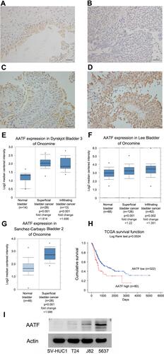
Figure 2 AATF positively regulates GC cell proliferation. (A) Transfection and siRNA knockdown efficiencies were confirmed by Western blot and RT-qPCR in T24 and 5637 cell lines, respectively. (B) CCK8 showed that growth rate was increased in T24 cells with AATF overexpression, while the cell growth rate was decreased in 5637 cells with AATF siRNA knockdown. (C) Colony formation assay demonstrated that colony counts were higher in T24 cells with AATF overexpression. AATF knockdown downregulated colony numbers formed by 5637 cell line. * p < 0.05.
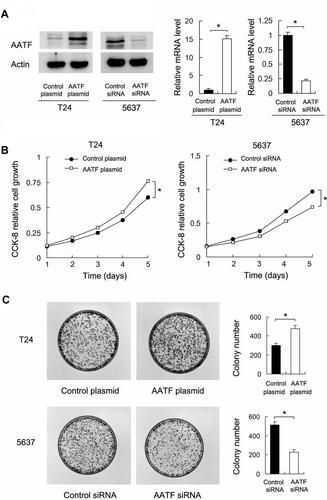
Figure 3 The effects of AATF on cell cycle and cisplatin inhibitory rate. (A) Cell cycle analysis showed that AATF overexpression upregulated S percentage in T24 cell line, while AATF siRNA decreased S percentage in 5637 cells. (B) The inhibitory rate of different cisplatin concentration in T24, T24/AATF, 5637, and 5637/AATFKD cells after cisplatin treatment. Ectopic AATF expression decreased inhibitory rate and increased IC50 in T24 cell line (IC50 con vs AATF: 12.4μM vs 14.5μM). AATF knockdown increased inhibitory rate and decreased IC50 value in 5637 cell line (IC50 con vs AATF KD: 12.3μM vs 10.1μM). *p < 0.05.

Figure 4 AATF regulates cisplatin sensitivity and apoptosis. (A) CCK8 cell viability assay showed that AATF overexpression increased cell viability while AATF knockdown decreased it after 24 and 48 h of cisplatin (10μM) treatment. The viability in cells without cisplatin was also shown. The fold changes of cell viability were indicated. (B) Annexin V/PI results showed that the cisplatin induced apoptosis rate was decreased in T24 cells transfected with AATF plasmid. AATF depletion upregulated cisplatin induced apoptosis in 5637 cell line. (C) JC-1 staining showed that ectopic AATF expression upregulated the mitochondrial membrane potential by decreasing green fluorescence percentage in T24 cells treated with cisplatin. AATF knockdown downregulated the mitochondrial membrane potential by increasing green fluorescence in 5637 cells treated with cisplatin. The changes of membrane potential in non-treated cells were not significant. *p < 0.05.
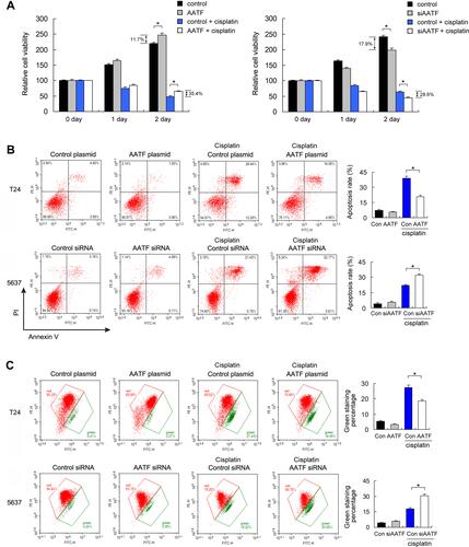
Figure 5 AATF regulates cyclin E and Survivin. (A) Western blotting showed that ectopic AATF expression decreased the levels of cytochrome c and cleaved PARP in cisplatin treated T24 cell line. AATF depletion increased the levels of cytochrome c and cleaved PARP in cisplatin treated 5637 cell line. (B) Western blotting and RT-qPCR showed ectopic AATF expression upregulated cyclin E and Survivin. AATF depletion downregulated cyclin E and Survivin at both mRNA and protein levels. (C) Analysis of TCGA data showed that AATF positively correlated with both cyclin E and Survivin mRNA in 408 cases of BC tissues. *p < 0.05.
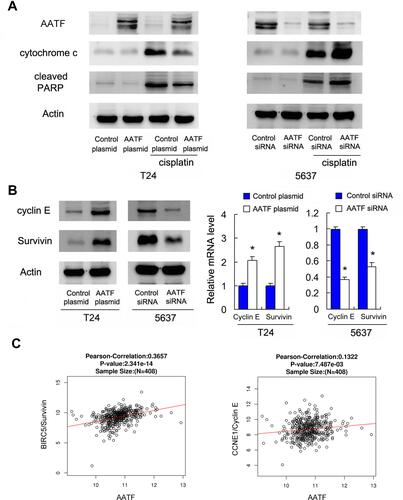
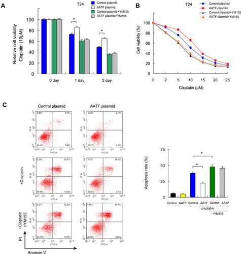
Figure 6 AATF regulates cisplatin-induced apoptosis through Survivin. (A) T24 cells with AATF overexpression were treated with Survivin inhibitor YM155 (10 μM) CCK-8 showed that AATF overexpression increased cell viability, which was abolished after treatment with YM155. Survivin inhibitor reversed the effect of AATF on cisplatin sensitivity. (B) The inhibitory rate of different cisplatin concentration in T24, T24/AATF, T24/YM155, and T24/AATF/YM155 cells after cisplatin treatment. T24 cells treated with YM155 also showed decreased IC50 and AATF overexpression could not significantly increased IC50 in YM155 treated cells. (C) Apoptosis assay showed that Survivin inhibitor YM155 upregulated cisplatin-induced apoptosis rate which was decreased by AATF. In T24 cells treated with YM155, the apoptosis rate was not changed significantly after AATF overexpression. *p < 0.05.