Figures & data
Table 1 Laboratory Testing Results
Table 2 IHC Staining for the Patient’s Tissue Samples
Table 3 Cases of Immune-Related Cystitis Following ICI: Clinical Characteristics and Malignancy Status
Figure 1 (A) Preoperative chest CT scan (captured on March 19th, 2018). (B) Histology revealed squamous carcinoma of the lung (x100). (C) Postoperative chest CT follow-up (captured on April 11th, 2018). (D) PET-CT scan (captured on July 18th, 2019). (E) Histology findings indicated SCLC. (F) PET-CT scan (captured on September 11th, 2019). Significant abnormal findings were noted (arrow).
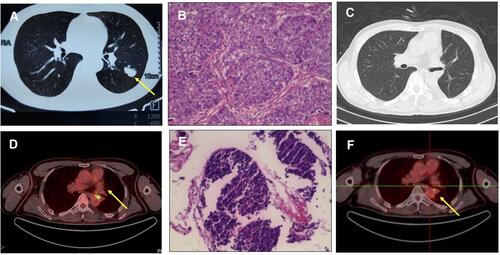
Figure 2 (A) Hepatic metastatic lesion and (B) bone metastases on PET-CT (captured on Nov. 11th, 2019). (C and D) Confirmation of CR after second line nivolumab and paclitaxel (captured on April 8th, 2020). (E) Cystoscopy showed that the entire bladder mucosa was edematous. (F) Histology of urothelium. Significant abnormal findings were noted (arrow).
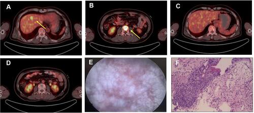
Figure 3 Radiotherapy planning to be delivered to bone metastases for this patient.
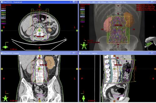
Figure 4 IHC staining for the patient’s tissue samples. (A–E) Lung squamous carcinoma staining for CD3, CD4, CD8, PD-1 and PD-L1, respectively. (F–J) Small cell lung cancer biopsy sample staining for CD3, CD4, CD8, PD-1 and PD-L1, respectively. (K–O) Urothelium biopsy staining for CD3, CD4, CD8, PD-1 and PD-L1, respectively.
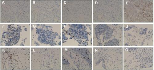
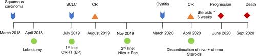
Figure 5 Clinical course of the patient. *means “times” (for 6 weeks).