Figures & data
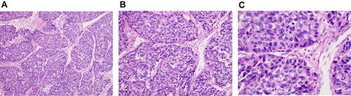
Figure 1 Biopsy specimen for tumor lesions.
Notes: Microscopic morphology of pancreatic hepatoid carcinoma. (A) at low power view, the poorly differentiated neoplasm, which showed hepatoid differentiation, had abundant sinusoids and cord-like or nest-like arrangement of tumor cells; (B) at medium power view, the tumor cells had abundant cytoplasm, large and hyperchromatic nuclei, and frequent mitosis; (C) higher magnification of the poorly differentiated carcinoma demonstrating probable evidence of hepatoid differentiation of tumor cells.