Figures & data
Figure 1 Spontaneous regression of metastases after the discontinuation of chemotherapy. Plain CT scans in cross-sectional views of lung metastases and retroperitoneal lymph node (RPLN) metastases at 1 month after chemotherapy discontinuation (A and B). Plain CT of lung metastases (C) and PET-CT of RPLN metastases (D) at 7 months after chemotherapy discontinuation. Both lung and RPLN metastases spontaneously regressed without further treatment. During this time, the patient’s hCG level decreased from 84,920 to 1402 IU/L.
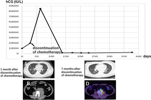
Figure 2 Clinical course of treatment with pembrolizumab. Since the patient’s hCG level started to re-increase after spontaneous regression continued for 8 months, immunotherapy with pembrolizumab was started. The hCG level decreased from 6500 to <1.0 IU/L after two doses of pembrolizumab. PET-CT 40 days after the start of treatment showed shrinkage of RPLN metastases with diminished metabolism.
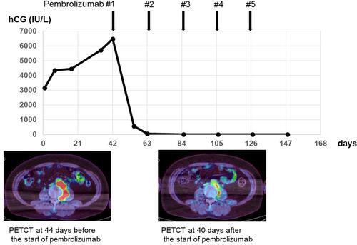
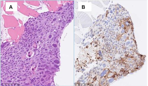
Figure 3 Pathological section of RPLN metastasis biopsy. The biopsy specimen showed the infiltration of mixed germ cell cancer. Immunohistochemistry revealed strong positive staining for PD-L1. (A) Hematoxylin and eosin staining. (B) VENTANA (SP-142) immunohistochemistry assay, magnification: 200x.