Figures & data
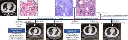
Figure 1 Timeline of the clinical course. Thoracic CT images of the tumor at baseline (A), after neoadjuvant treatment with gefitinib (C and D), and at postoperative follow-up (G and H); Hematoxylin-Eosin stained slide of the core needle biopsy (B), resected primary lesion (E) and resected lymph node (F).
Abbreviations: NSE, neuron-specific enolase; SCC-Ag, squamous cell carcinoma antigen; CYFRA-21-1, cytokeratin 19 fragment; CEA, carcinoembryonic antigen.