Figures & data
Table 1 Cerebrospinal Fluid Test Showed That the Anti-γ-Aminobutyric Acid Type B Receptor (GABAB) Antibody IgG Was Positive
Figure 1 (A1 and A2) The baseline CT scan of the patient’s chest in August 2020. (B1 and B2) CT scan of the chest after two cycles of durvalumab in October 2020.
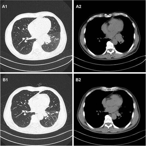
Figure 2 (A1 and A2) The baseline MRI scan of the patient’s head in August 2020. (B1 and B2) MRI scan of the head after the first seizure in October 2020. (C1 and C2) MRI scan of the head after the second seizure.
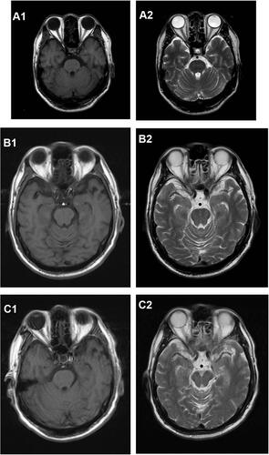
Figure 3 (A1 and A2) The result of the lump in the left lung pathological histopathology and immunohistochemistry by local bronchoscopy biopsy performed showed that is small-cell endocrine cancer.
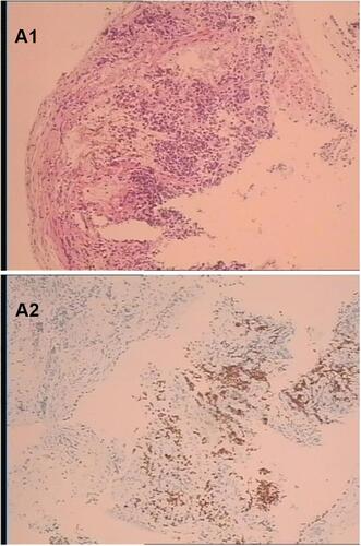
Figure 4 (A1 and A2) PETCT on August 27, 2020; The increased mass of FDG metabolism in the hyperplastic block indicated by the arrow.
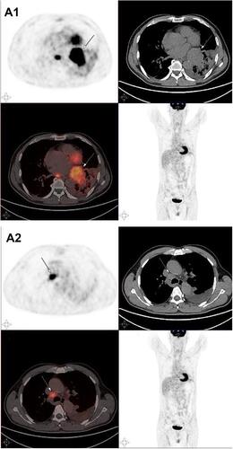
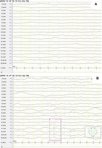
Figure 5 (A) The patient’s EEG after the first seizure in October 2020. (B) The patient’s EEG after the second seizure in October 2020. The pink box showed EEG becomes abnormal situation with sharp and slow waves.