Figures & data
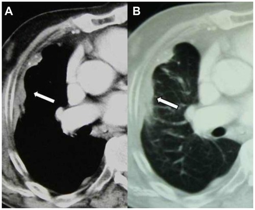
Figure 1 Axial computed tomography images of the right lung in the (A) mediastinal and (B) pulmonary window demonstrate the pleural mass corresponding to mesothelioma (initial examination). antigen, and thyroid transcription factor-1. Metastasis in the adrenal gland remained stable. Bone scans and brain CT scans were not pathological. No confirmation was made on the malignancy of the adrenal gland. Pleural thickness at diagnosis was 90 mm × 20 mm, and 40 mm × 15 mm at the end of the first-line treatment. Follow-up has continued for 9 years; the disease is stable without any disease progression.
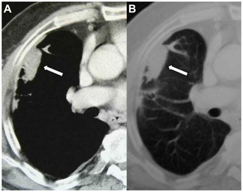
Figure 2 Axial computed tomography images of the right lung in the (A) mediastinal and (B) pulmonary window 9 years after initial examination show that the bulky side of the mesothelioma has slightly decreased.