Figures & data
Table 1 Primer
Figure 1 TSC22D2 expression in CRC cell lines predicted poor survival. (A) Expression of TSC22D2 in tumor and non-tumor tissues in TCGA database. (B and C) mRNA and protein expression of TSC22D2 in CRC cells. (D) Immunofluorescent images show the TSC22D2 protein primarily localized to the cell cytoplasm. (E and F) TSC22D2 expression and CRC patients’ clinical prognosis.
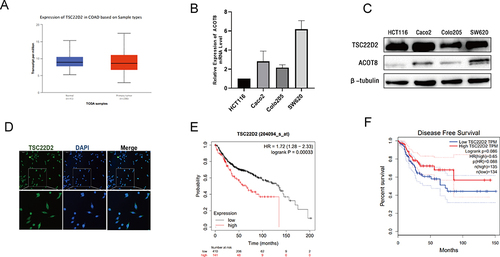
Figure 2 TSC22D2 inhibits the proliferation, migration and invasion of CRC cells. (A) HCT116 cells were transfected with control si-RNA (si-NC) or TSC22D2 si-RNA and subjected to Western blot for TSC22D2expression. (B and C). CCK8 assay and Colony assay verified that TSC22D2 knockdown enhanced the capacity of proliferative and colony formation of HCT116 cells. (D). The number of migrating and invading HCT116 cells were increased by TSC22D2 know down. (E–H). SW620 cells transfected with pcDNA/TSC22D2 or empty vector and measured by Western blot for TSC22D2 expression. Proliferation and migration capability of cells were also assessed. *P < 0.05.
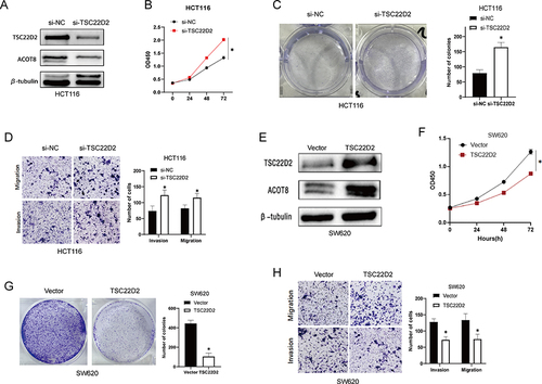
Figure 3 TSC22D2 altered the expression of EMT-related proteins. (A and B) E-cadherin, Vimentin, N-cadherin were detected by Western blot in HCT116 cells and SW620 cells transfected with si-RNA or TSC22D2 plasmid. *P < 0.05.
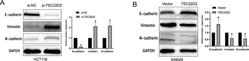
Figure 4 TSC22D2 inhibits CRC growth in mice. (A). Subcutaneous tumorigenesis in mice was evaluated after PPP1R14B knockdown. (B and C) Both tumor volume and tumor weight in TSC22D2 knockdown group (n=5) were obviously higher than those in control group (n=6). *P < 0.05.
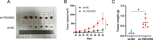
Figure 5 Identification of TSC22D2-interacting proteins using co-IP/MS. (A). There were 8 overlapping TSC22D2 binding proteins identified. (B). Correlation of TSC22D2 with ACOT8 in the GEPIA database. (C). ACOT8 was immunoprecipitated by TSC22D2 in HCT116 cells. (D). Expression of ACOT8 mRNA in tumor and non-tumor tissues in TCGA database.
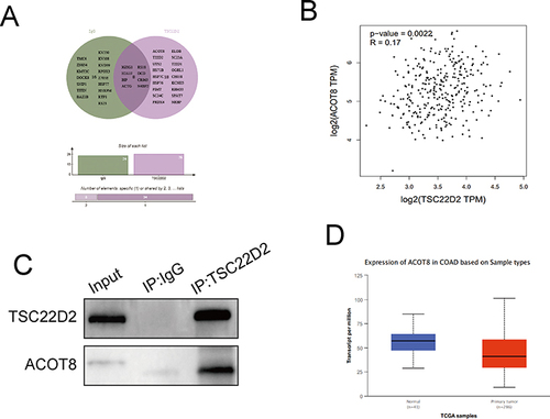
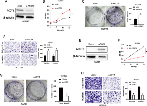
Figure 6 ACOT8 inhibits the proliferation, migration and invasion of CRC cells. (A). HCT116 cells were transfected with control si-RNA (si-NC) or ACOT8 si-RNA and subjected to Western blot for TSC22D2 expression. (B and C). CCK8 assay and Colony assay verified that ACOT8 knockdown enhanced the capacity of proliferative and colony formation of HCT116 cells. (D). The number of migrating and invading HCT116 cells were increased by ACOT8 know down. (E–H). SW620 cells transfected with pcDNA/ACOT8 or empty vector and measured by Western blot for ACOT8 expression. Proliferation and migration capability of cells were also assessed. *P < 0.05.