Figures & data
Figure 1 (A) The terminal ileum shows no obvious occupancy. (B) Preoperative CT image showing the morphology of the end of the descending colon–sigmoid colon–rectum intestinal tube thickening with multifocal areas of exudation and small lymph nodes. There were slightly hyperdense foci in the terminal sigmoido-rectal mesentery of the descending colon.(The descending colon tumour is indicated by the red arrow in the figure).
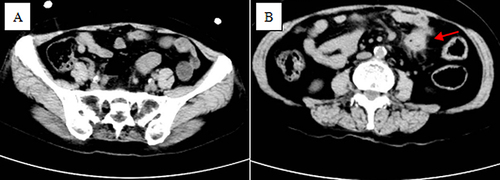
Figure 2 (A) Gross pathology of a tumour of the terminal ileum. (B) Gross pathology of a tumour of the descending colon.
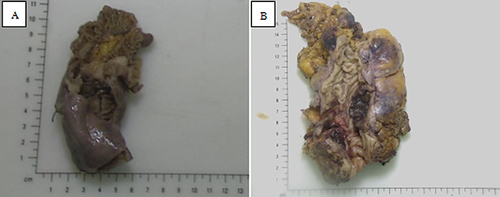
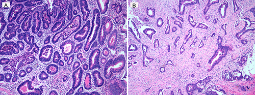
Figure 3 (A) The picture shows ileal adenocarcinoma cells found in sections of the resected tissue. (B) The picture shows the descending colon intestinal adenocarcinoma cells found in the resected tissue sections.