
Figures & data
Figure 1 Radiographic findings before and after dacomitinib treatment. (A) Chest X-ray showed a mass-like opacity in the left lung field. (B) CT scan displayed a 7×7 cm mass in the left upper lobe, with enlarged contralateral mediastinal lymph nodes and evidence of multiple bony metastases. (C) Chest X-ray and (D) CT scan revealed a reduced lung tumor size in the left upper lobe after dacomitinib therapy.
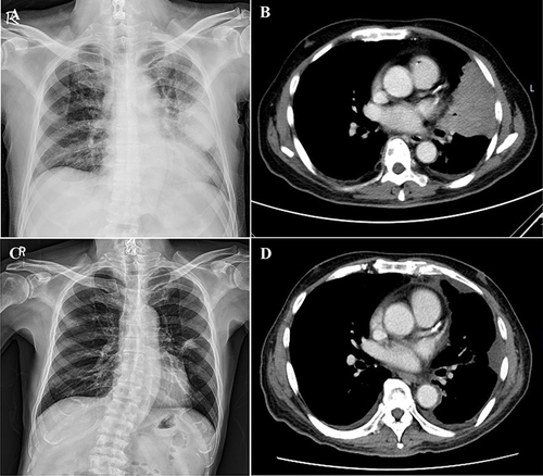
Figure 2 MRI scans displayed a metastatic lesion in the left thalamus before and after dacomitinib treatment. (A) Axial MRI scan showed a 2.2×1.6 x 1.5 cm lesion with mixed signal intensity (arrow), suggestive of a hemorrhagic metastatic lesion. (B) Axial MRI scan revealed complete remission of the previously noted metastatic lesion.
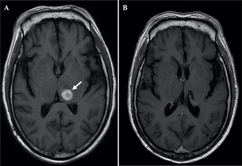
Figure 3 Radiographic findings before and after chemotherapy treatment. (A) Chest X-ray and (B) CT scan demonstrated an enlarged primary lesion in the left upper lobe with associated pleural effusion. (C) Chest X-ray and (D) CT scan revealed a decreased size of the tumor in the left upper lobe with diminished pleural effusion after chemotherapy.
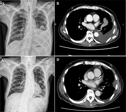
Figure 4 Diagram of the CCDC6-RET fusion gene on chromosome 10. The schematic illustrates the fusion between exon 1 of the CCDC6 gene and exon 12 of the RET gene, resulting in the CCDC6-RET fusion gene.
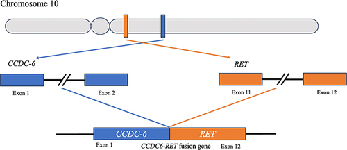
Figure 5 Brain CT scans depicting hydrocephalus. (A) Exhibited a dilated lateral ventricle (arrow). (B) Showed an expanded 3rd ventricle (arrow).
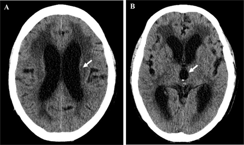
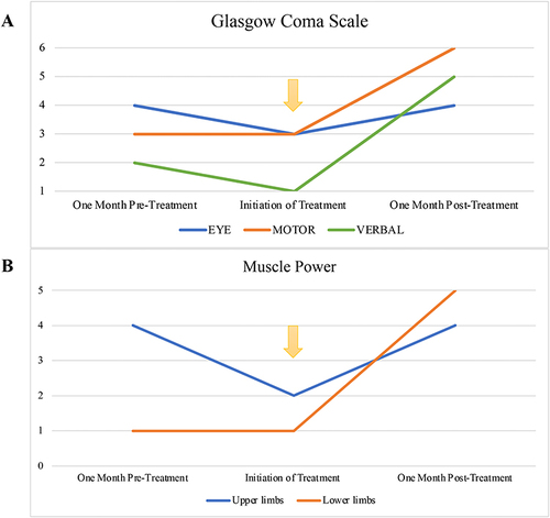
Figure 6 Clinical progression measured by Glasgow Coma Scale and muscle strength evaluation. (A) Displayed improvement in Eye, Motor, and Verbal responses as per the Glasgow Coma Scale. (B) Illustrated the increase in muscle power for both upper and lower limbs. The initiation of combined treatment is denoted by a yellow arrow.
Data Sharing Statement
All data related to the study are included in the paper.