Figures & data
Figure 1 Surgical specimen of paraganglioma of vagina.
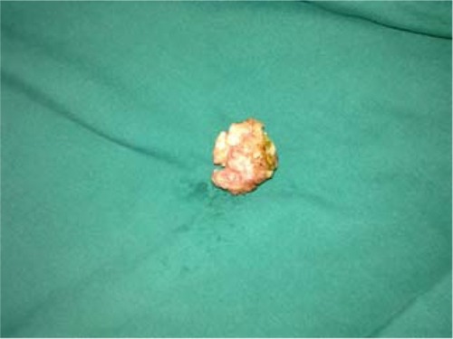
Figure 2 Vaginal tumor cells.
Note: Vaginal tumor cells showed nested aggregation and microscopically small nests composed of neoplastic cells having abundant cytoplasms demarcated by delicate fibrous stroma and capillaries in the Zellballen pattern (hematoxylin and eosin staining, 100×).
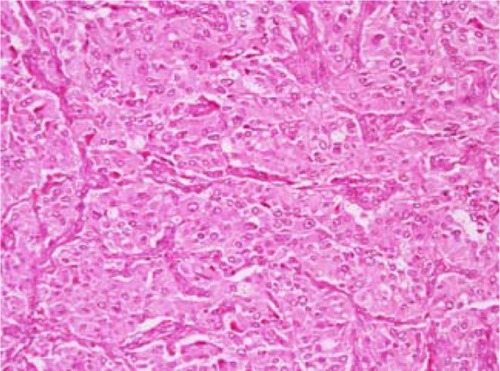
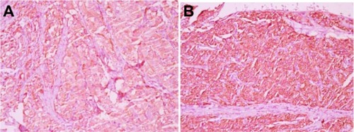
Figure 3 Positive immunohistochemical staining for chromogranin (400×) (A); positive immunohistochemical staining for synaptophysin (400×) (B).