
Figures & data
Table 1 Patient characteristics
Table 2 Results of administration of heat shock protein 70
Figure 1 Case report of intratumoral Hsp70 therapy in patient 5.
Notes: (A) MRI brain images in a 4-year-old boy with a choroid plexus carcinoma before intratumoral injections of Hsp70 (upper panels) with MRI 4 weeks after the last intralesional infusion of Hsp70. (B) Analysis of cytokine (IL-1β, IL-1Ra, IL-2, IL-4, IL-6, interferon-gamma, tumor necrosis factor-alpha) levels in cerebrospinal fluid after a single Hsp70 infusion. Cerebrospinal fluid was collected 3, 6, 12, 24 and 48 hours after injection of chaperone and cytokines were detected by enzyme-linked immunosorbent assay. (C) Immunofluoresence image of choroid plexus carcinoma section, with Hsp70 detected by monoclonal antibodies BRM-22a (with Alexa568 conjugated secondary antibodies, red color). Nuclei were stained by DAPI (blue color). Scale bar, 75 μm. On the right is a magnified image of tumor cells on the immunohistochemistry assay with BRM-22a antibodies to Hsp70 revealed by standard biotin-peroxidase reaction (nuclei stained by hematoxylin). Scale bar, 10 μm.
Abbreviations: h, hours; Hsp70, heat shock protein 70; TNFα, tumor necrosis factor alpha; IL-1Ra, interleukin-1 receptor antagonist; IFNγ, interferon gamma; DAPI, 4’,6-diamidino-2-phenylindole; IL, interleukin; MRI, magnetic resonance imaging.
Abbreviations: h, hours; Hsp70, heat shock protein 70; TNFα, tumor necrosis factor alpha; IL-1Ra, interleukin-1 receptor antagonist; IFNγ, interferon gamma; DAPI, 4’,6-diamidino-2-phenylindole; IL, interleukin; MRI, magnetic resonance imaging.
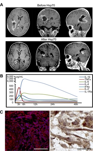
Table 3 Main parameters in peripheral blood lymphocyte subpopulations
Table 4 Main parameters in cytokine levels in peripheral blood