Figures & data
Figure 1 Schematic representation of experimental protocols performed in this study.
Notes: Tg(k18:dsred) zebrafish were intraperitoneally injected either with buffer only (mock-control group, n=6) or with buffer containing 40 μg/g of Lipo-Dox (Lipo-Dox™-injected group, n=12, #1–#12). We injected Lipo-Dox one dose (40 μg/g) at day 0, and subsequently injected one dose per week for 4 weeks (days 7, 14, 21, and 28), and examined the zebrafish phenotypic defects at each check point (days 10, 17, 24, 31, 38, 45, and 51).
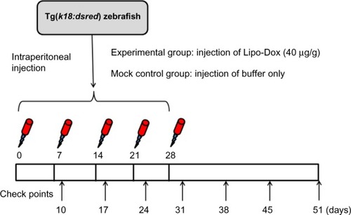
Figure 2 Abdominal hemorrhage and fin necrosis are observed in zebrafish embryos after Lipo-Dox™ injection.
Notes: (A–E) Mock control. (A′–E′) Tg(k18:dsred) zebrafish (#2) was injected with buffer containing 40 μg/g of Lipo-Dox.
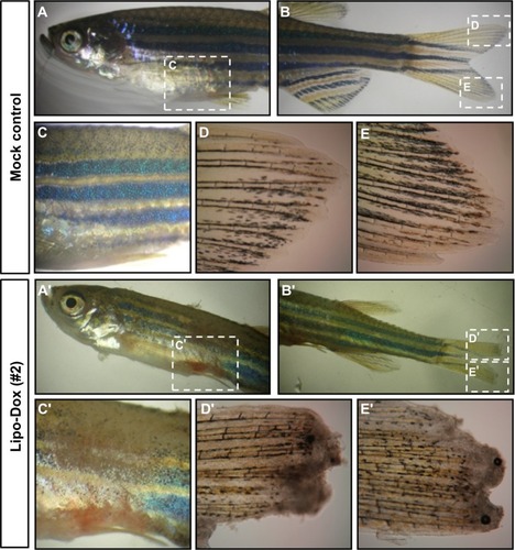
Figure 3 Effects of Lipo-Dox™ on the zebrafish’s caudal fin (I) and epidermis (II).
Notes: Lipo-Dox-injected Tg(k18:dsred) zebrafish (#5 and #6) were observed under microscopy with bright field (A-I–F-I) or an RFP filter (A-II–F-II). Fin necrosis (arrow indicates) and keratinocyte dissociation are the evident phenotypes in the Lipo-Dox-injected zebrafish.
Abbreviation: RFP, red fluorescent protein.
Abbreviation: RFP, red fluorescent protein.
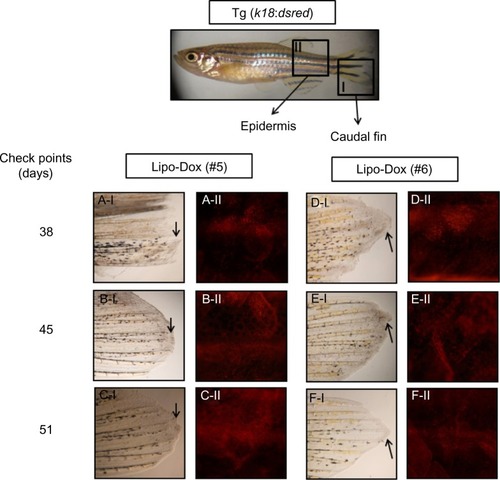
Figure 4 Histological examination of Lipo-Dox™-injected embryos.
Notes: Fish derived from the mock control (A–C) or Lipo-Dox-injected groups (D–F) were transverse sectioned and stained with hematoxylin/eosin Y. (A and C) Lateral side; (B and E) Ventral region; (C and F) Intestine. Yellow star indicates the position of goblet cell. Black arrows indicate the positions of epidermis lesions.
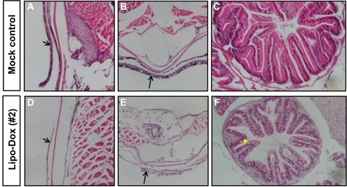
Table 1 Generalized linear regression based on the GEE method for assessing the effect of Lipo-Dox™ on the weight growth rates of embryos
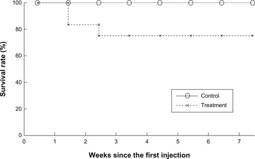
Figure 5 Kaplan–Meier estimates of survival curves for the mock control and Lipo-Dox™-injected groups.