Figures & data
Figure 1 T1-weighted contrast enhancement magnetic resonance imaging scan of Case 1.
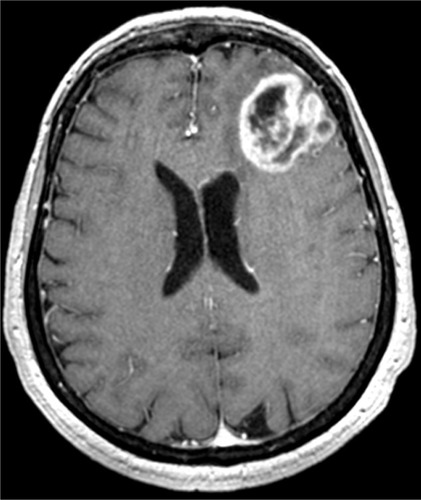
Table 1 List of primary antibodies used for immunohistochemistry
Table 2 List of primary antibodies used for immunofluorescence
Figure 2 Immunohistochemistry.
Notes: Case 1: (A) perivascular cell arrangement; H&E, 10×; (B) necrosis and polymorphic cells; H&E, 10×; (C) GFAP-positive perivascular cells; DAB, 10×; (D) Ki-67/MIB-1 LI; DAB, 20×. Case 2 (first intervention): (E) aspect of oligodendroglioma; H&E, 10×; (F) GFAP-negative palisades; DAB, 10×; (G) rhythms and palisades; H&E, 10×; (H) Ki-67/MIB-1 LI; DAB, 20×. Case 2 (second intervention): (I) perivascular arrangement of astrocytic cells; H&E, 20×; (J) id, H&E; 20×; (K) GFAP-positive perivascular cells; DAB, 20×; (L) Ki-67/MIB-1 LI; DAB, 10×. Case 3 (M) perivascular arrangement of large cells; H&E, 20×; (N) polymorphic cells and vessel hyalinization; H&E, 10×; (O) GFAP-positive perivascular cells; DAB, 20×; (P) Ki-67/MIB-1 LI; DAB, 20×.
Abbreviations: DAB, 3,3’-Diaminobenzidine; H&E, hematoxylin and eosin; LI, labeling index; id, perivascular arrangement of astrocytic cells.
Abbreviations: DAB, 3,3’-Diaminobenzidine; H&E, hematoxylin and eosin; LI, labeling index; id, perivascular arrangement of astrocytic cells.
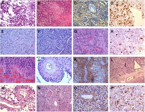
Figure 3 Sanger direct sequencing.
Notes: Case 1: (A) electropherograms for the stop mutation c.822G>A (p.Trp274*) in PTEN exon 8 in lymphocytes, primary tumor, neurospheres, and adherent cells; (B) electropherograms for the missense mutation c.824G>A (p.Cys275Tyr) in TP53 exon 8 in lymphocytes, primary tumor, neurospheres, and adherent cells. Case 2 (first intervention): (C) electropherograms for the splice variant c.209 + 1G>T in PTEN intron 3 in lymphocytes, primary tumor, and adherent cells.
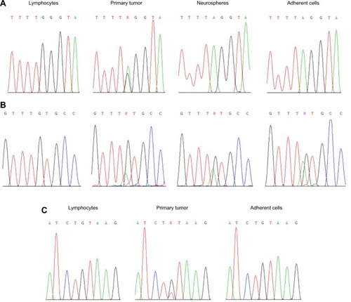
Table 3 Genetic and epigenetic alterations in tumors and matched cell lines
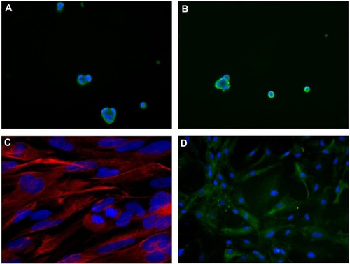
Figure 4 Immunofluorescence.
Notes: Case 1: (A) neurospheres, Musashi-1 expression; 40×; (B) id, nestin expression; 40×; (C) adherent cells, GFAP expression; 40× (confocal microscopy). Case 2 (first intervention): (D) adherent cells, GFAP expression; 40×. DAPI counterstaining was used for all.
Abbreviations: DAPI, 4′,6-diamidino-2′-phenylindole dihydrochloride; id, neurospheres.
Abbreviations: DAPI, 4′,6-diamidino-2′-phenylindole dihydrochloride; id, neurospheres.