Figures & data
Figure 1 CT scan of the case of HCC spontaneous regression.
Notes: CT scan showed massive-type liver tumors located in the right lobe. They had a nonuniform low-density in Couinaud’s segment 7 and 8.
Abbreviations: CT, computed tomography; HCC, hepatocellular carcinoma.
Abbreviations: CT, computed tomography; HCC, hepatocellular carcinoma.
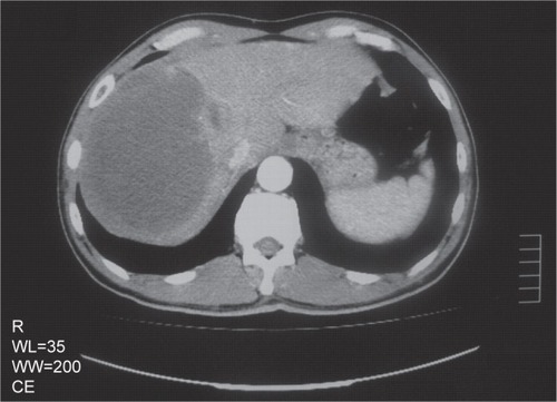
Figure 2 Macroscopical features of the case of HCC spontaneous regression.
Notes: The tumor on the cut surface, measuring 10×9 cm, was yellowish and brown with a partially fibrous capsule and septum-like structures. Moreover, a reddish-brown nodule less than 1.2 cm was identified as surviving malignant tumor.
Abbreviation: HCC, hepatocellular carcinoma.
Abbreviation: HCC, hepatocellular carcinoma.
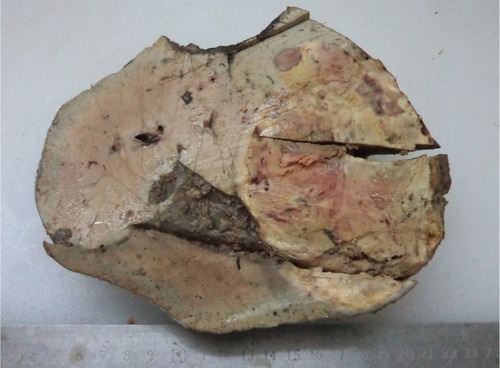
Figure 3 Microscopical features of the case of HCC spontaneous regression.
Notes: (A) Histologic examination shows coagulation necrosis of most of the zone in the tumor, and ghosts of the tumor cells are arranged in a trabecular pattern. (B) The surviving malignant zone was about 11.6 mm at low magnification. (C) At high magnification, the surviving part of the tumor was composed of highly pleomorphic cells arranged in sheets and trabeculae. Tumor cells are large and polygonal with central, vesicular nuclei and prominent nucleoli. Concurrently, some inflammatory cells infiltrate between tumor cells. The macrophages are the predominant inflammatory cells, with abundant cytoplasm and large nuclei (marked with the green arrows). (D) Many inflammatory cells infiltrated the periphery of tumor. Lymphocytes, neutrophils, and macrophages were detected.
Abbreviation: HCC, hepatocellular carcinoma.
Abbreviation: HCC, hepatocellular carcinoma.
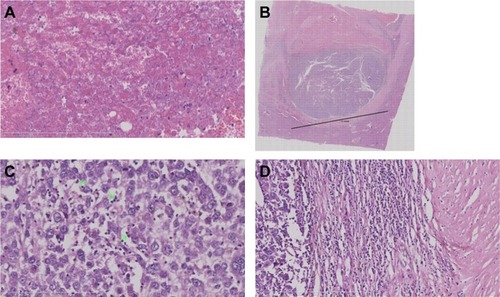
Figure 4 Immunological features of the case of HCC spontaneous regression.
Notes: (A) CD68 demonstrated individual positive cells in the central zone of the surviving portion of the tumor. (B) CD68 demonstrated an amount of positive cells in the fibrous capsule of the periphery of the tumor. The left part of the image is the tumor. The right part of the image is the periphery of the tumor. (C) CD163 demonstrated more positive cells with large size and abundant cytoplasm in the central zone of partially surviving tumors. (D) CD163 demonstrated many positive cells in the fibrous capsule of the periphery of the tumor. However, the positive cells in the fibrous capsule were smaller than in partially surviving tumors. The left part of the image is of partially surviving tumors. The right part of the image is the periphery of tumor. (E) CD163 demonstrated a small amount of positive cells in peritumoral normal liver tissue.
Abbreviation: HCC, hepatocellular carcinoma.
Abbreviation: HCC, hepatocellular carcinoma.
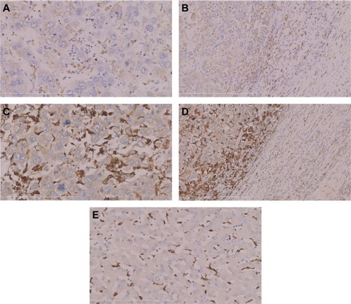
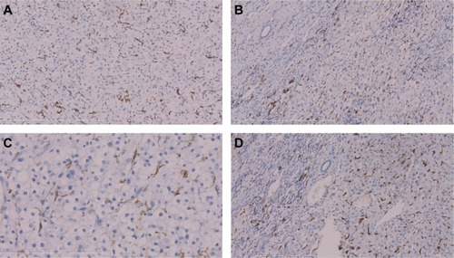
Figure 5 Immunological features of non-specific HCC as a control.
Notes: (A) CD68 demonstrated many positive cells in non-specific HCC, and (B) in the periphery of tumor. (C) CD163 demonstrated many positive cells in non-specific HCC, and (D) in the periphery of tumor, and the morphology of positive cells was similar. The density and size of both CD68+ and CD163+ cells were similar in non-specific HCC.
Abbreviation: HCC, hepatocellular carcinoma.
Abbreviation: HCC, hepatocellular carcinoma.