
Figures & data
Figure 1 Brain axial magnetic resonance imaging T1-weighted sequence after gadolinium intravenous administration showing thickening and enhancement of bilateral vestibulocochlear nerves and solitary lesion in right cerebellar hemisphere.
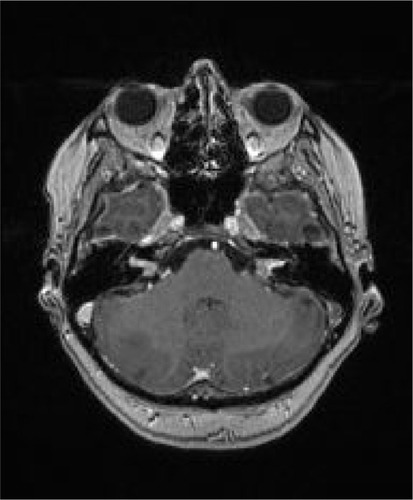
Figure 2 Entire stomach wall infiltrated with solid nests of tumor tissue, with occasional central necrosis. Tumor is adenocarcinoma grade III.
Notes: Hematoxylin and eosin stain; ×200 magnification.
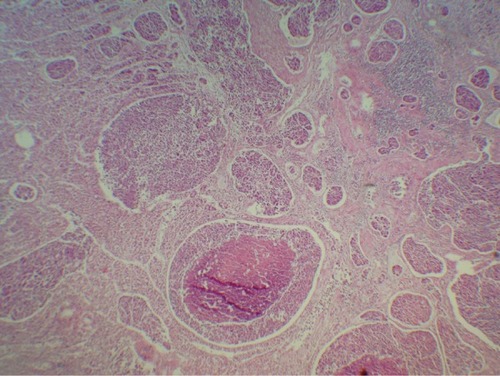
Table 1 Bilateral hearing loss as presenting symptom of gastric cancer
Table 2 Threshold cycle values by quantitative real-time polymerase chain reaction