
Figures & data
Figure 1 Image registration by automatic bone matching and manual fine-tuning method in head.
Notes: (A) The cross section, coronal plane, and sagittal plane in an NPC patient’s planning CT, and we use nasal septum as a bony landmark for automatic bone matching and manual fine-tuning method (the yellow frame represents registration area); (B) the cross section of the head in planning CT image; (C) the cross section of the head in cone-beam CT image; (D) a typical fusion image with planning CT. The areas circled in red indicate the primary gross volume (GTVnx).
Abbreviations: NPC, nasopharyngeal carcinoma; CT, computed tomography.
Abbreviations: NPC, nasopharyngeal carcinoma; CT, computed tomography.
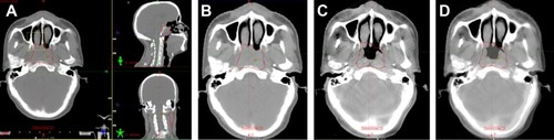
Figure 2 Image registration by automatic bone matching and manual fine-tuning method in upper neck.
Notes: (A) The cross section, coronal plane, and sagittal plane in an NPC patient’s planning CT, and we use 1–3 cervical vertebrae as a bony landmark for automatic bone matching and manual fine-tuning method (the yellow frame represents registration area); (B) the cross section of upper neck in planning CT image; (C) the cross section of upper neck in cone-beam CT image; (D) a typical fusion image with planning CT. The areas circled in red indicate the involved lymph nodes (GTVnd).
Abbreviations: NPC, nasopharyngeal carcinoma; CT, computed tomography.
Abbreviations: NPC, nasopharyngeal carcinoma; CT, computed tomography.
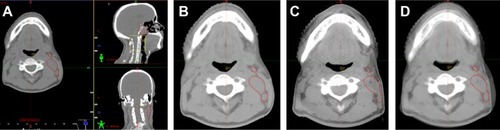
Figure 3 Image registration by automatic bone matching and manual fine-tuning method in lower neck.
Notes: (A) The cross section, coronal plane, and sagittal plane in an NPC patient’s planning CT, and we use 4–6 cervical vertebrae as a bony landmark for automatic bone matching and manual fine-tuning method (the yellow frame represents registration area); (B) the cross section of lower neck in planning CT image; (C) the cross section of lower neck in cone-beam CT image; (D) a typical fusion image with planning CT. The areas circled in red indicate the involved lymph nodes (GTVnd).
Abbreviations: NPC, nasopharyngeal carcinoma; CT, computed tomography.
Abbreviations: NPC, nasopharyngeal carcinoma; CT, computed tomography.
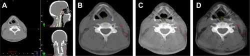
Figure 4 Setup errors of 30 patients in the (A) right–left direction; (B) superior–inferior direction, and (C) anterior–posterior direction (the mean ± SD).
Abbreviations: RL, right–left; SI, superior–inferior; AP, anterior–posterior; SD, standard deviation.
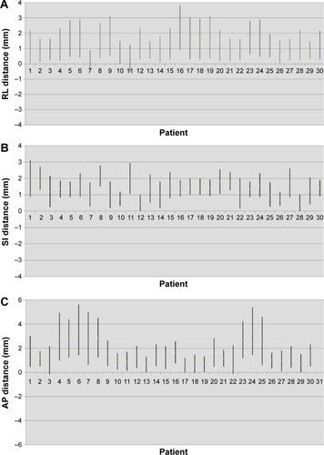
Table 1 The translational shifts >3 mm in RL, SI, and AP directions of head, upper neck, and lower neck
Table 2 Summary of interfraction translational error (mm) in each dimension
Table 3 Analysis of variance of setup errors in different parts in RL, SI, and AP directions