
Figures & data
Figure 1 Abdominal enhanced magnetic resonance imaging with T1 findings.
Notes: (A) An 8×6 cm mass in the head of the pancreas (white arrow). (B) Liver metastasis from mixed acinar-endocrine carcinoma of the pancreas (red arrow).
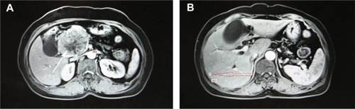
Figure 2 Histopathology of the pancreas tumor.
Notes: (A) (a) Hematoxylin–eosin image of the pancreas tumor (HEX200). (b) Immunohistochemistry of trypsin (×400, Abcam). (c) Immunohistochemistry of chymotrypsin (×400, Abcam). (d) Immunohistochemistry of Ki-67 (×400, Abcam [Cambridge, MA, USA]). (B) (a) Immunohistochemistry of chromogranin A(×400, Abcam) (b) Immunohistochemistry of synaptophysin (×400, Abcam). (c) Immunohistochemistry of CD56 (×400, Abcam). (d) Immunohistochemistry of neuron-specific enolase (NSE) (×400, Abcam).
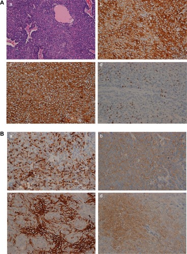
Figure 3 Histopathology of the liver metastasis.
Notes: (A) (a) Hematoxylin–eosin image of the liver metastasis (HEX100). Left is adjacent cancerous tissue and right is the liver metastasis. (b) Immunohistochemistry of hepatocyte paraffin 1 (Hep Par 1) (×100, Dako). Adjacent cancerous tissue was positive for immunohistochemistry of Hep Par 1 and the liver metastasis was negative for the same. (c) The liver metastasis was negative for immunohistochemistry of Hep Par 1 (×400, Dako Denmark A/S, Glostrup, Denmark). (d) Immunohistochemistry of CD56 (×400, Abcam). (B) (a) Immunohistochemistry of trypsin (×400, Abcam), indicating that the tissue had an acinar component. (b) Immunohistochemistry of chromogranin A(×400, Abcam). (c) Immunohistochemistry of synaptophysin (×400, Abcam). (d) Immunohistochemistry of neuron-specific enolase (NSE) (×400, Abcam). Chromogranin A, synaptophysin, and NSEwere weakly positive for immunohistochemistry, suggesting that the tissue of the liver metastasis had endocrine component.
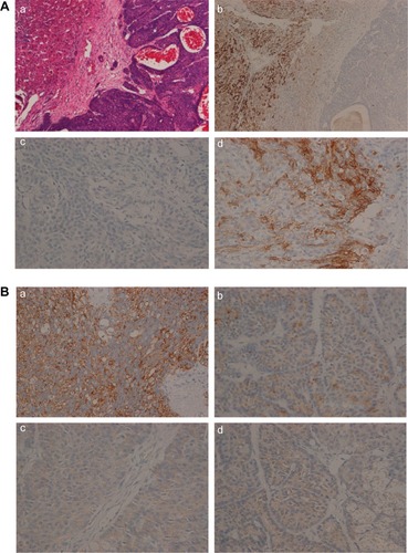
Figure S1 Survival curve for the patients after surgery.
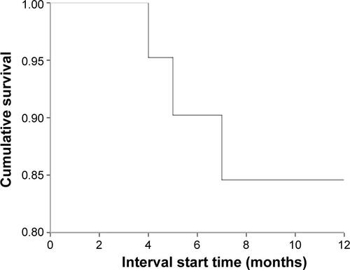
Table S1 Reported cases of mixed acinar-endocrine carcinoma in pancreas
Table S2 Life table showing survival time after surgery (months)Table Footnotea