
Figures & data
Table 1 Comparison between VR and MIP for reconstruction of perigastric arteries (case)
Figure 1 VR and MIP reconstruction techniques for perigastric arteries.
Notes: (A) VR reconstruction technique for perigastric arteries. The figure shows the gastric arteries of a 66-year-old man diagnosed with gastric cancer of the gastric corpus. (B) VR reconstruction technique for perigastric arteries. The representative figure is from a 58-year-old man diagnosed with early gastric cancer located at the cardia of stomach. (C) MIP reconstruction technique for perigastric arteries from the same patient in (A). (D) MIP reconstruction technique for perigastric arteries from the same patient in (B).
Abbreviations: CA, celiac axis; CHA, common hepatic artery; LGA, left gastric artery; SA, splenic artery; PHA, proper hepatic artery; SMA, superior mesenteric artery; RGeA, right gastroepiploic artery; RHA, right hepatic artery; RGeA, right gastroepiploic artery; VR, volume rendering; MIP, maximum intensity projection.
Abbreviations: CA, celiac axis; CHA, common hepatic artery; LGA, left gastric artery; SA, splenic artery; PHA, proper hepatic artery; SMA, superior mesenteric artery; RGeA, right gastroepiploic artery; RHA, right hepatic artery; RGeA, right gastroepiploic artery; VR, volume rendering; MIP, maximum intensity projection.
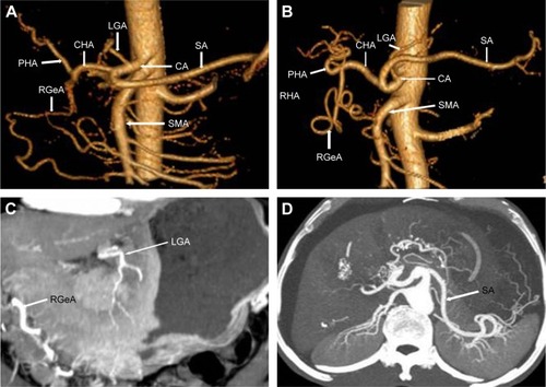
Table 2 Comparison for perigastric arterial types by angiography (%)
Figure 2 Three-dimensional reconstruction of gastric vessel anatomy and rare variations.
Notes: (A) CHA originating from superior mesenteric artery. This is an image of the gastric arteries of a 69-year-old woman diagnosed with gastric cancer located at gastric antrum. Hepatic artery arising from mesenteric artery could be regarded as arterial variation Type II. (B) CHA originating from superior mesenteric artery of a 73-year-old man diagnosed with gastric cancer of gastric antrum. Hepatic artery arising from mesenteric artery could be regarded as arterial variation Type II. The variant vessles LGA in (A), and CHA in (B) are written in red.
Abbreviations: CHA, common hepatic artery; SA, splenic artery; LGA, left gastric artery; RGeA, right gatroepiploic artery; SMA, superior mesenteric artery; PHA, proper hepatic artery.
Abbreviations: CHA, common hepatic artery; SA, splenic artery; LGA, left gastric artery; RGeA, right gatroepiploic artery; SMA, superior mesenteric artery; PHA, proper hepatic artery.
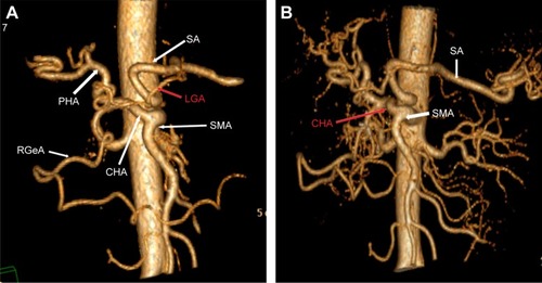
Figure 3 Right hepatic artery deriving from GDA.
Notes: (A) The image shows the gastric arteries of a 58-year-old man diagnosed with early gastric cancer at the gastric corpus. Right hepatic artery was originating from GDA, which could to be classified as Type III. (B) Right hepatic artery deriving from GDA. The figure is from a 61-year-old man diagnosed with gastric cancer located at gastric antrum. Right hepatic artery was originating from GDA, which could be classified as Type III. The variant vessles SMA in (A), and GDA in (B) are written in red.
Abbreviations: CHA, common hepatic artery; SA, splenic artery; RHA, right hepatic artery; SMA, superior mesenteric artery; RGeA, right gastroepiploic artery; GDA, gastroduodenal artery; LHA, left hepatic artery.
Abbreviations: CHA, common hepatic artery; SA, splenic artery; RHA, right hepatic artery; SMA, superior mesenteric artery; RGeA, right gastroepiploic artery; GDA, gastroduodenal artery; LHA, left hepatic artery.
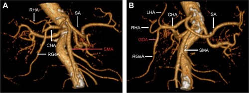
Figure 4 LGA originating from aorta.
Notes: This was from a 77-year-old man diagnosed with gastric cancer located at gastric antrum. LGA (in red) and superior mesenteric artery were directly originating from aorta.
Abbreviations: CHA, common hepatic artery; SA, splenic artery; SMA, superior mesenteric artery; RGeA, right gastroepiploic artery; PHA, proper hepatic artery; LGA, left gastric artery.
Abbreviations: CHA, common hepatic artery; SA, splenic artery; SMA, superior mesenteric artery; RGeA, right gastroepiploic artery; PHA, proper hepatic artery; LGA, left gastric artery.
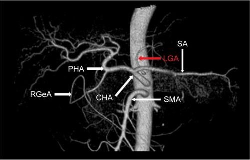
Table 3 Comparison of arterial variations between our group and Hiatt’s (case)