Figures & data
Figure 1 Cytotoxicity of LDM or LDM combined with IDO on HepG2 cells determined via an MTT assay.
Note: Data represented triple results, mean ± SD.
Abbreviations: LDM, lidamycin; IDO, iodized oil; SD, standard deviation.
Abbreviations: LDM, lidamycin; IDO, iodized oil; SD, standard deviation.
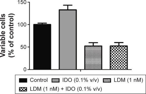
Figure 2 DSA and ultrasound images of a VX2 tumor transplanted in a rabbit liver.
Notes: (A) Angiography of the hepatic artery showing a rich blood supply to the tumor, especially the rim of the tumor. (B) Embolization with IDO, which was deposited well in the lesion. (C) Ultrasound image showing a relative hyperintensity VX2 tumor in the liver lobe (white arrow). The (+) and (−) labels were used to measure the size of the tumor, with the distance between the (+) and (−) labels giving the diameter.
Abbreviations: DSA, digital subtraction angiography; IDO, iodized oil.
Abbreviations: DSA, digital subtraction angiography; IDO, iodized oil.
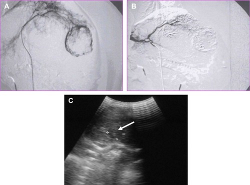
Table 1 Inhibitory efficacy of ADM + IDO and LDM + IDO on rabbit VX2 tumors
Figure 3 Changes in the rabbit serum ALT and AST levels after TOCE.
Notes: Blood chemical analysis suggested that there was an increase in the serum AST (A) and ALT (B) levels in the ADM + IDO and LDM + IDO groups at 3 days after perfusion as compared to those at 0 and 10 days, but there was no significant difference between the ADM + IDO and LDM + IDO groups. The results indicated that the influence of ADM + IDO or LDM + IDO perfusion on rabbit serum ALT and AST levels was transient and recoverable.
Abbreviations: ALT, alanine aminotransferase; AST, aspartate aminotransferase; TOCE, transarterial oily chemoembolization; ADM, Adriamycin; IDO, iodized oil; LDM, lidamycin.
Abbreviations: ALT, alanine aminotransferase; AST, aspartate aminotransferase; TOCE, transarterial oily chemoembolization; ADM, Adriamycin; IDO, iodized oil; LDM, lidamycin.
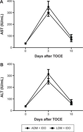
Figure 4 Western blot analysis of HIF-1α expression in rabbit liver tissue.
Notes: (A) is the western blot of HIF-1α and (B) was the quantitative analysis of A using the Image J software (developed by the National Institutes of Health). Control represents the HIF-1α expression in normal rabbit liver tissue, while ADM + IDO and LDM + IDO represent the HIF-1α expression in VX2 tumor tissues that were collected at day 3 after TOCE with ADM + IDO or LDM + IDO, respectively. There was a significant difference between the ADM + IDO and LDM + IDO groups compared with the control group. The data indicated that HIF-1α was upregulated in rabbit VX2 tumor tissue after TOCE. *P<0.05, compared to the control.
Abbreviations: HIF-1α, hypoxia-inducible factor-1α; ADM, Adriamycin; IDO, iodized oil; LDM, lidamycin; TOCE, transarterial oily chemoembolization.
Abbreviations: HIF-1α, hypoxia-inducible factor-1α; ADM, Adriamycin; IDO, iodized oil; LDM, lidamycin; TOCE, transarterial oily chemoembolization.
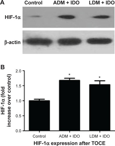
Figure 5 Immunohistochemistry analysis of VX2 tumor sections.
Notes: Effects of ADM + IDO and LDM + IDO on tumor proliferation assessed via PCNA levels, tumor microvessel density assessed via CD31 levels, and tumor angiogenesis assessed via VEGF levels in paraffin-embedded VX2 tumor sections. The IHC analysis of PCNA, CD31, and VEGF expression indicated the enhanced inhibition of cell proliferation and angiogenesis in the LDM + IDO group compared to the ADM + IDO group. *P<0.05.
Abbreviations: ADM, Adriamycin; IDO, iodized oil; LDM, lidamycin; PCNA, proliferating cell nuclear antigen; CD31, cluster of differentiation 31; VEGF, vascular endothelial growth factor; IHC, immunohistochemistry.
Abbreviations: ADM, Adriamycin; IDO, iodized oil; LDM, lidamycin; PCNA, proliferating cell nuclear antigen; CD31, cluster of differentiation 31; VEGF, vascular endothelial growth factor; IHC, immunohistochemistry.
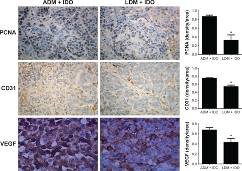
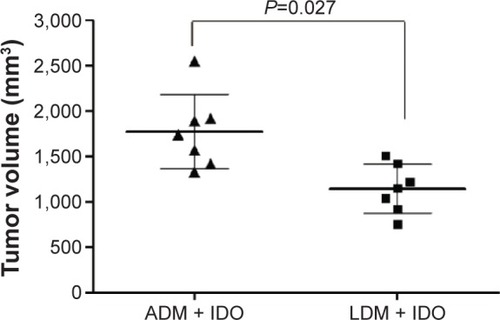
Figure 6 Volume of tumor following treatment with ADM + IDO or LDM + IDO at day 10 after TOCE.
Note:
P=0.027 for the LDM + IDO group compared with the ADM + IDO group.
Abbreviations: ADM, Adriamycin; IDO, iodized oil; LDM, lidamycin; TOCE, transarterial oily chemoembolization.
Abbreviations: ADM, Adriamycin; IDO, iodized oil; LDM, lidamycin; TOCE, transarterial oily chemoembolization.