
Figures & data
Table 1 Patient characteristics (N=133)
Table 2 List of assays used and the respective sample dilutions
Table 3 List of used primary and secondary antibodies, their manufacturers, and incubation conditions
Table 4 List of antigens and cellular proteins in tumor cell lysates
Table 5 List of antigen/danger patterns in tumor cell lysates selected as typical indicators for renal clear cell carcinoma (frequency of occurrence >70%, n>10)
Figure 1 Pattern of antigen distribution in different RCCs: frequencies of occurrence were calculated for all RCC samples and stratified by sex.
Abbreviations: RCC, renal clear cell carcinoma; pos, positive; neg, negative.
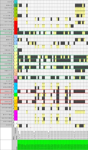
Table 6 Absolute values of different tumor-associated antigens and cellular marker proteins (CYFRA, NSE, TPA, and TPS) in nontumor tissue (normal) and the respective tumor tissue (ccRCC) (ND, not determined)
Table 7 Absolute values of carbonic anhydrase IX (CA-IX) in nontumor tissue and the respective tumor tissue
Figure 2 (A) Overlay immune-stains for an antibody panel in tumor tissue (examples): CD10 (purple), CD11b (green), CD21 (blue), CD4 (light green), CD40 (cyan), CD45RA (yellow), CD45RO (violet), CD8 (pink), CA IX (red), and propidium iodine (mint); (B) Overlay immune-stains for an antibody panel in single cell suspension (examples): NSE (red), p53 (green), and CD105 (blue), combined signals: NSE + p53 + CD105 (white), p53 + CD105 (yellow), and NSE + CD105 (violet); (C) Overlay immune stains for an antibody panel in tumor tissue for CA-IX (green), NSE (pink), and CXCR4 (blue); and (D) Immune stain for a NSE-specific antibody (white dots) in nontumor kidney tissue (negative control).
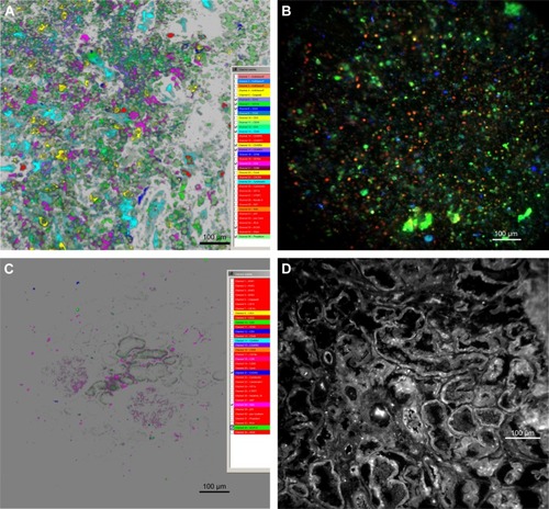
Table 8 Detection of a humoral immune response in mice (n=28) after two intradermal shots (200 μL) with a TCL; total protein and antigen levels in the respective TCL are given (missing values justified by sample availability)