Figures & data
Figure 1 miR-34b expression in NPC tissue samples and cell lines.
Notes: (A) Relative miR-34b expression levels in NPC and adjacent peritumoral tissues. miR-34b displayed a decreased expression level in NPC tissue compared with the adjacent peritumoral tissue (**P<0.01). (B) Relative miR-34b expression levels in various cell lines as determined by qRT-PCR analysis (**P<0.01).
Abbreviations: NPC, nasopharyngeal carcinoma; qRT-PCR, quantitative reverse transcription-polymerase chain reaction.
Abbreviations: NPC, nasopharyngeal carcinoma; qRT-PCR, quantitative reverse transcription-polymerase chain reaction.
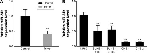
Figure 2 Ectopic overexpression of miR-34b suppressed the proliferation of SUNE-6-10B cells and induced G0/G1 cell cycle arrest in vitro.
Notes: (A) The expression levels of miR-34b in SUNE-6-10B cell lines were verified after transfection with 100 nM miR-34b mimics and miR-control. (B) The effect of miR-34b on cell proliferation was measured using the MTT assay in SUNE-6-10B cell lines (*P<0.05). (C and D) Flow cytometric analysis of the cell cycle distribution of SUNE-6-10B cells transiently transfected with 100 nM miR-34b mimics and the control. (E) The expression levels of P21, CyclinD, CyclinE, Bcl-2, Bax, and Caspase-3 were measured by Western blot analysis. GAPDH was used as an internal control. Each independent experiment was repeated three times. All the experiments were performed in triplicate, and the results were analyzed by ANOVA and are expressed as mean ± SD (**P<0.01).
Abbreviations: MTT, 3-(4,5-dimethyl-2-thiazolyl)-2,5-diphenyl-2H-tetrazolium bromide; GAPDH, glyceraldehyde 3-phosphate dehydrogenase; ANOVA, analysis of variance; OD, optical density.
Abbreviations: MTT, 3-(4,5-dimethyl-2-thiazolyl)-2,5-diphenyl-2H-tetrazolium bromide; GAPDH, glyceraldehyde 3-phosphate dehydrogenase; ANOVA, analysis of variance; OD, optical density.
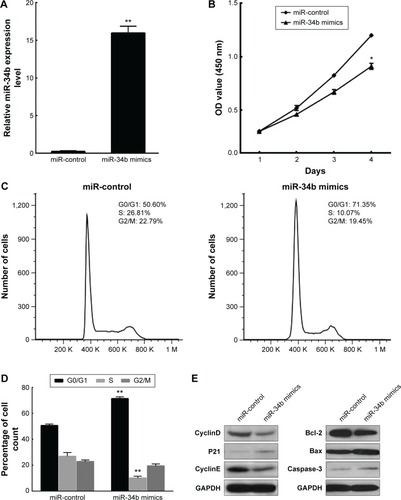
Figure 3 miR-34b targets the 3′-UTR of the USP22 gene.
Notes: (A) Schematic graph of the putative binding sites of miR-34b in the USP22 3′-UTR. Mut 3′-UTR indicates the USP223′-UTR with a mutation in the miR-34b binding sites. (B) miR-34b mimics downregulated the activity of a luciferase reporter containing wild-type USP22 3′-UTR (**P<0.01) but not of a reporter containing mutant USP22 3′-UTR. (C) Forty-eight hours after miR-34b mimic and control transfection of SUNE-6-10B cells, the mRNA level of USP22 decreased significantly compared with the miR-control, as determined by qRT-PCR (**P<0.01). (D) Forty-eight hours after miR-34b mimic and control transfection of SUNE-6-10B cells, the protein level of USP22 decreased significantly compared with the miR-control, as determined by Western blot analysis. GAPDH was used as an internal control. Each independent experiment was repeated three times. All the experiments were performed in triplicate. The results were analyzed by ANOVA and are expressed as mean ± SD.
Abbreviations: 3′-UTR, 3′-untranslated region; USP22, ubiquitin-specific peptidase 22; qRT-PCR, quantitative reverse transcription-polymerase chain reaction; GAPDH, glyceraldehyde 3-phosphate dehydrogenase; ANOVA, analysis of variance; wt, wild type; Ploy A, polyadenylic acid; CMV, Cytomegalovirus.
Abbreviations: 3′-UTR, 3′-untranslated region; USP22, ubiquitin-specific peptidase 22; qRT-PCR, quantitative reverse transcription-polymerase chain reaction; GAPDH, glyceraldehyde 3-phosphate dehydrogenase; ANOVA, analysis of variance; wt, wild type; Ploy A, polyadenylic acid; CMV, Cytomegalovirus.
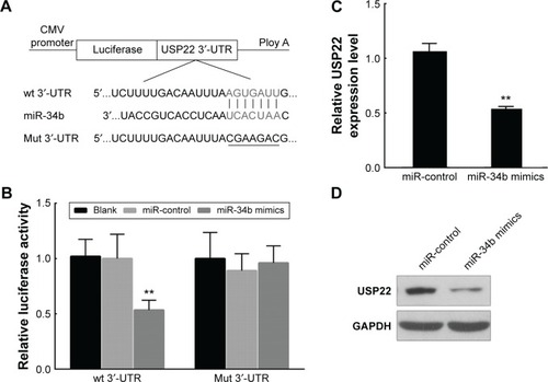
Figure 4 USP22 regulates the proliferation and cell cycle of SUNE-6-10B cells in vitro.
Notes: (A) Quantitative RT-PCR analysis of USP22 mRNA expression in nasopharyngeal carcinoma tissue and noncancerous nasopharyngeal mucosa. (B) Western blot analysis of USP22 in various NPC cell lines. (C) The abundance of USP22 proteins was assessed in SUNE-6-10B cells by Western blot analysis following ectopic overexpression of USP22. (D) The effect of USP22 overexpression on cell proliferation was measured using the MTT assay in SUNE-6-10B cell lines. (E and F) Flow cytometric analysis of the cell cycle distribution of SUNE-6-10B cells transiently transfected with USP22-overexpressing plasmid or control plasmid. (G) The abundance of USP22 proteins in SUNE-6-10B cells after siRNA transfection was analyzed by Western blot analysis. (H) The effect of siRNA-USP22 on cell proliferation in SUNE-6-10B cell lines was measured using the MTT assay. (I and J) Flow cytometric analysis of the cell cycle distribution of SUNE-6-10B cells that were transiently transfected with 100 nM siRNA-USP22 or the control. Each independent experiment was repeated three times. All the experiments were performed in triplicate, and the results were analyzed by ANOVA and are expressed as mean ± SD (**P<0.01; *P<0.05).
Abbreviations: USP22, ubiquitin-specific peptidase 22; RT-PCR, reverse transcription-polymerase chain reaction; NPC, nasopharyngeal carcinoma; MTT, 3-(4,5-dimethyl-2-thiazolyl)-2,5-diphenyl-2H-tetrazolium bromide; siRNA, small interfering RNA; ANOVA, analysis of variance; GAPDH, glyceraldehyde 3-phosphate dehydrogenase; OD, optical density.
Abbreviations: USP22, ubiquitin-specific peptidase 22; RT-PCR, reverse transcription-polymerase chain reaction; NPC, nasopharyngeal carcinoma; MTT, 3-(4,5-dimethyl-2-thiazolyl)-2,5-diphenyl-2H-tetrazolium bromide; siRNA, small interfering RNA; ANOVA, analysis of variance; GAPDH, glyceraldehyde 3-phosphate dehydrogenase; OD, optical density.
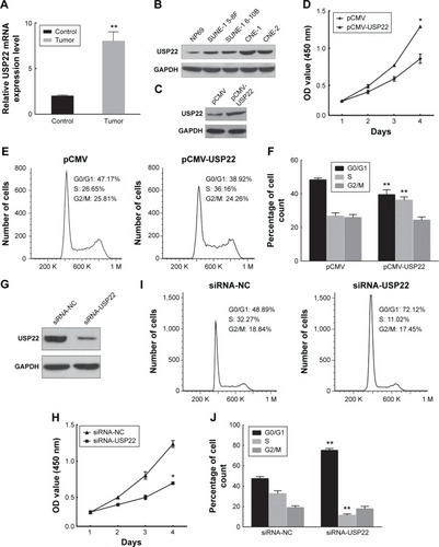
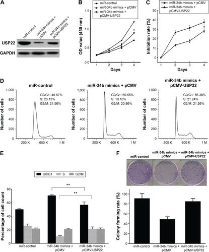
Figure 5 miR-34b represses USP22 to suppress SUNE-6-10B growth.
Notes: (A) Western blot analyses were performed to evaluate the efficiency of miR-34b overexpression and USP22 reintroduction in SUNE-6-10B cells. (B and C) The cell proliferation activity was analyzed using the MTT assay. (D and E) Flow cytometric analysis of the cell cycle distribution of the indicated SUNE-6-10B cells. (F) Colony formation ability was analyzed by colony formation assay. **P<0.01; *P<0.05.
Abbreviations: USP22, ubiquitin-specific peptidase 22; MTT, 3-(4,5-dimethyl-2-thiazolyl)-2,5-diphenyl-2H-tetrazolium bromide; GAPDH, glyceraldehyde 3-phosphate dehydrogenase; OD, optical density.
Abbreviations: USP22, ubiquitin-specific peptidase 22; MTT, 3-(4,5-dimethyl-2-thiazolyl)-2,5-diphenyl-2H-tetrazolium bromide; GAPDH, glyceraldehyde 3-phosphate dehydrogenase; OD, optical density.