Figures & data
Figure 1 Standard of care increases Myxoma virus (MYXV) spread.
Notes: (A) Timeline of treatment protocol for all experiments. (B) Cells were plated in replicates onto 12-well plates. Percent of GFP+ cells were analyzed by flow cytometry 12 hours after infection with vGFP and treatment as indicated in the protocol. Data represent summation of three independent experiments (P<0.05). (C) Cells were plated into replicates on to 12-well plates. The numbers of replication competent MYXV particles were determined by titering after harvesting from cells pretreated with the indicated treatment protocols in duplicate. Data represent the summation of two independent experiments. (D) Cells were plated onto 6-well plates in replicates. Images of GFP positive foci, infected with vGFP and treated as indicated, at 4× magnification 72 hours after infection under fluorescent microscopy. (E) Mean foci area was assessed at 24-hour intervals after infection with vGFP and treated as indicated for 72 hours. There was a significant difference between cells treated with 1 Gy of radiation and untreated cells (*=P<0.011). Data are representative of three independent experiments.
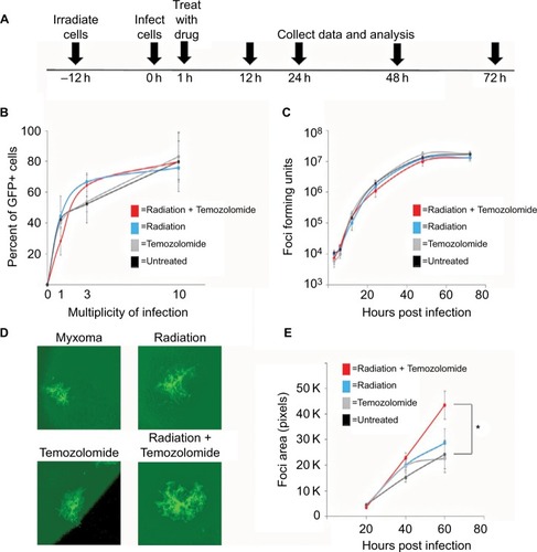
Figure 2 Infection of primary GBM biopsies.
Notes: Biopsies were collected from three patients with GBM. Samples were prepared via the standard Stoppini Slice Culture as outlined. Samples were then treated as indicated and infected with vGFP for 48 hours prior to imaging at a magnification of 4×. Images taken via fluorescent microscopy.
Abbreviations: GBM, glioblastoma multiforme; MYVX, Myxoma virus.
Abbreviations: GBM, glioblastoma multiforme; MYVX, Myxoma virus.
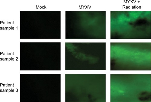
Figure 3 In vitro analysis of cell viability of combined treatment conditions.
Notes: (A) Cells were plated in replicates and treated as indicated prior to infection with vGFP. Cell viability of U118 tumor cells measured by MTT assay as percent of control at 72 hours postinfection. All treatments resulted in significant decrease in viability compared to untreated cells (P<0.001, individual values not shown). Combination therapy of chemotherapeutic drug, MXYV and radiation significantly reduced cell viability compared to all other treatment groups at 72 hours (***<0.001). Data are the summation of three independent experiments. (B) Cells were plated in replicates and treated as indicated prior to infection with vGFP. Fold increase of Caspase-3 activity compared to control was measured using a commercially available kit, as described, at 24-hour intervals postinfection. Combination treatment of chemotherapeutic drug, MXYV and radiation significantly increased Caspase-3 activity compared to any other treatment group at 72 hours (***<0.001, **<0.01). Data are the summation of three independent experiments. (C) Western blot analysis of p-Akt, Caspase-3 and actin in GL261 mouse brain tumor cells, T9 rat brain tumor cells and U118 human tumor cells. Samples were treated with each chemotherapeutic agent either with or without radiation and MYXV.
Abbreviations: MYXV, Myxoma virus; Rad, radiation;.
Abbreviations: MYXV, Myxoma virus; Rad, radiation;.
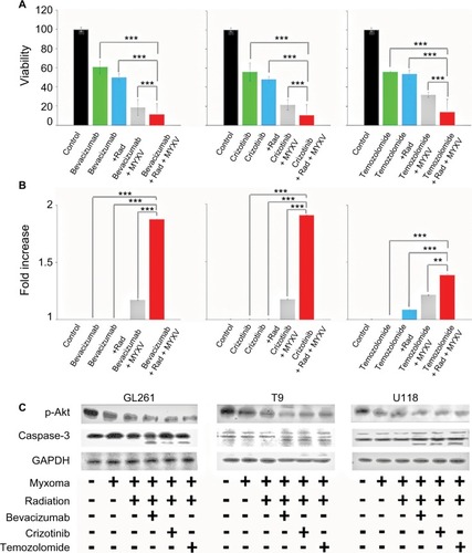
Figure 4 Ex vivo analysis of cell viability of combined treatment conditions.
Notes: (A) Patient biopsies were harvested, cultured in duplicate 96-well plates, infected with vGFP and treated as indicated. Cell viability of patient biopsies was measured by MTT assay as described at 24-hour intervals postinfection. Addition of all treatments resulted in a significant decrease in cell viability (P<0.01, individual values not shown). Combination treatment of chemotherapeutic agent, radiation and MYXV showed a significant decrease in cell viability compared to any other treatment combination (***<0.001, **<0.01, *<0.05). Data are the summation of three independent experiments. (B) Patient biopsies were harvested, cultured in duplicate 96-well plates, infected with vGFP and treated as indicated. Caspase-3 activity of patient biopsies was measured by a colorimetric kit, as described, at 24-hour intervals postinfection. Fold increase of Caspase-3 activity was calculated as compared to control. Addition of all treatments resulted in a significant increase in Caspase-3 activity (P<0.01, individual values not shown). Combination treatment of chemotherapeutic agent, radiation and MYXV showed a significant increase in Caspase-3 activity compared to any other treatment combination (***<0.001, **<0.01). Data are the summation of three independent experiments. (C) Western blot analysis of p-Akt, Caspase-3 and actin in three patient GBM biopsies. Samples were treated with each chemotherapeutic agent either with or without radiation and MYXV.
Abbreviations: GBM, Glioblastoma; MYXV, Myxoma virus; Rad, radiation; MYXV, Myxoma virus; GBM, Glioblastoma.
Abbreviations: GBM, Glioblastoma; MYXV, Myxoma virus; Rad, radiation; MYXV, Myxoma virus; GBM, Glioblastoma.
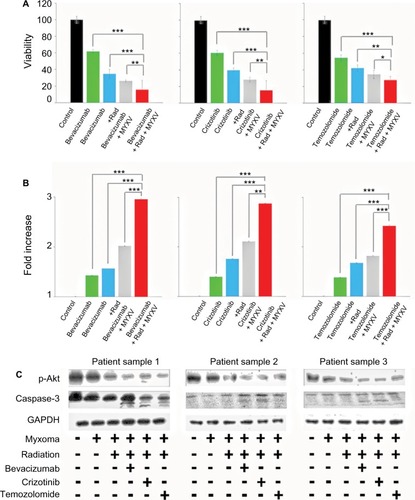
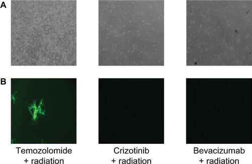
Figure S1 (A) Images of U118 cells treated with the corresponding treatment viewed under phase at 72 hours after infection. (B) GFP positive foci of infected U118 cells treated with the corresponding treatment regimen 72 hours after infection.