Figures & data
Figure 1 (A) Pedigree of patient’s family with inherited CCDC40 pathogenic variants. Black arrow, proband. Half-colored symbol, heterozygous CCDC40 variant carrier. Solid symbol, patient (affected). (B) Lung high-resolution computed tomography of the patient showed diffuse nodules among small airway and bronchiectasis in right middle lung and both lower lungs. (C) High-resolution computed tomography of the patient showed sinusitis.
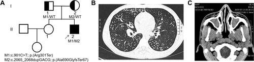
Figure 2 (A) Sanger-sequencing chromatograms and co-segregation analysis for patient’s family. Mutation 1 (NM_017950.3:c.901C>T, p.(Arg301*)) and mutation 2 (NM_017950.3:c.2065_2068dup, p. (Ala690Glyfs*67)) were identified in patient’s family. (B) Expression levels of CCDC40 are verified using RT-qPCR. A significantly reduced expression of CCDC40 is observed in the peripheral blood from the patient. (C and D) Location of CCDC40 mutations identified in this study are shown with arrows in the gene and protein. Predicted protein domains (coiled coil domains) are indicated by rectangles.
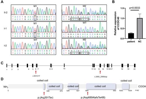
Figure 3 (A) Ciliary beating patterns of healthy cilia and our patient with CCDC40 mutations. (B) Papanicolaou staining showing morphology of the sperms from the patient demonstrated short, bent, coiled, and irregular flagella and other MMAF phenotypes. Scale bar, 10μm. (C) Scanning electron microscope of the sperms from the patient, showing bent, short, coiled and irregular flagella. Scale bar, 5μm.
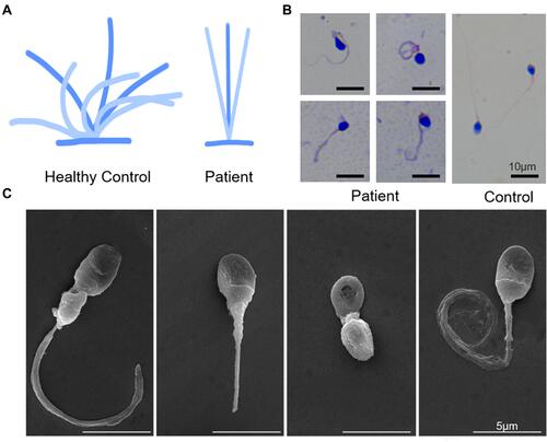
Table 1 Semen Parameters and Sperm Flagella Morphology in the Patient Carrying CCDC40 Variants and the Normal Control
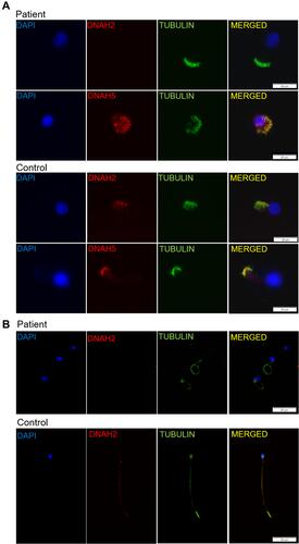
Figure 4 (A) Immunofluorescence analysis of nasal cilia cell of the patient and healthy control, showing presence of outer dynein arm protein DNAH5 and absence of the inner dynein arm protein DNAH2 in the patient. Scale bar, 20μm. (B) Immunofluorescence staining of sperms from the patient and the healthy control with α-tubulin antibodies (green) and DNAH2 (red), a marker for MMAF phenotype. DNAH2 is absent in the sperms from the patient. Scale bar, 20μm.