Figures & data
Figure 1 Clinical features of the patient: dark, thickened and tightened lamellar scales spread over almost all the skin surface with hair loss and ectropion.
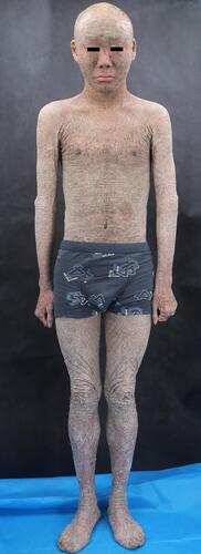
Figure 2 Dermoscopic features of the patient. Dermoscopy (×50 magnification) shows alopecia and pustules on his head (A), greasy light-brown scales on his face (B), “sheet-like” scales on his hand (C), pre-thoracic skin (D) as well as “bark-like” thickened scales with deep fissures between scales on the abdominal area (E) and on his thigh (F).
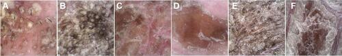
Figure 3 Histology (hematoxylin and eosin, ×100 magnification) shows severe lamellar hyperkeratosis, mild hypertrophy of the spinous layer, and vacuolated degeneration of some spinous cells. In the stratum corneum, there were some cross-sections of hair shaft (arrows). One of those structures was highlighted and magnified in the upper right corner (red box).
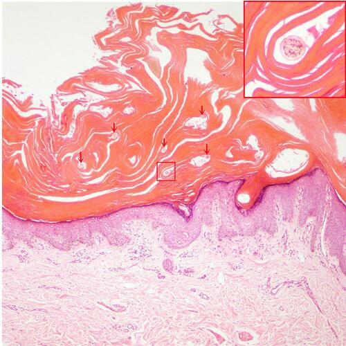
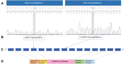
Figure 4 TGM1 mutations. (A) and (B) TGM1 mutations in the proband. (C) TGM1 structure. TGM1 has 15 exons and the translation start is located in the second exon.Citation26 (D) Protein domains of TGM1. It consists of five chains (817 amino acids): anchoring domain (1–92), β-sandwich domain (94–246), catalytic domain (247–572), β-barrel 1 domain (573–688), and β-barrel-2 domain (689–817).Citation19